

 полная версия
A Manual of the Operations of Surgery
Vascular adhesions to the wall which require many ligatures certainly add to the dangers of the case, while adhesions to the anterior wall of the abdomen render the operation, especially its first stages, much more difficult, preventing the cyst from being recognised.
2. The condition of the pedicle is of great importance. If it is too short, it prevents the use of the clamp, as if applied it is apt either to pull the uterus up, or, pulling the clamp down, to make undue traction on the wound, and rupture any adhesions. This is especially the case where much flatus is generated, or where the patient is naturally stout.
Treatment.—Where the pedicle is just long enough to allow the clamp to be applied, and yet too short to leave room for any distension of the abdomen without undue tension, the best plan is to transfix it with a stout double thread just below the clamp, tie it in two halves, and bring the threads out past the clamp, so that, if tension does occur, the clamp may be removed, the part beyond it cut off, and the rest allowed to slip back into the pelvis, the ligatures being kept out at the mouth of the wound.
Or again, it is sometimes possible, after applying one clamp firmly as near the tumour as possible, to apply another above it when the greater part of the tumour has been cut away; when the second is firmly fixed it may then be safe to remove the first, and thus an artificially elongated pedicle is obtained.
When still shorter, two plans remain for selection—(1.) to transfix the pedicle in one or more points, then, securing it in two, three, or more portions, cut it off above the ligatures and return it, leaving the ligatures at the lower end of the wound. This gives a free drain for pus, but theoretically the sloughing pedicle might be expected to set up peritonitis; (2.) to transfix and tie the pedicle with one or more loops of stout string, cut the ends off short, and return the whole affair, closing the external wound at once. Theoretically there are grave objections to this plan, but it has proved very successful, especially in the hands of Dr. Tyler Smith.
Another ingenious modification, sometimes useful in a short narrow pedicle, is to tie it as close to the cyst as possible, bring the ligature out at the wound, and then with a strong harelip needle transfix the pedicle, along with both sides of the wound, just below the ligature.
When the pedicle is excessively broad and stout, it should be transfixed by strong needles and double threads in various places, and thus tied in several portions. Absence of the pedicle greatly adds to the danger in any given case. Various plans have been tried, as cutting the attachment through slowly by the écraseur, ligature of each vessel separately, so many as twelve being sometimes required, and cauterising the stump. The latter, as used by Mr. Baker Brown, has met with a large measure of success, and is much used now.143
Dr. Keith for a time operated with antiseptic precautions, but has now (1883) entirely given up the use of the spray, which he believes has especial dangers in abdominal surgery.
Operation for Strangulated Inguinal Hernia.—The great rule to be remembered with regard to this, as well as all other operations for hernia, is, that the earlier it is performed the better chance the patient has. Once a fair trial has been given to the taxis, aided by proper position of the patient, the warm bath, and specially chloroform, the operation should be performed.
The patient should be placed on his back with his shoulders elevated, and the knee of the affected side slightly bent. The groin should then be shaved, and the shape and size of the tumour, with the position of the inguinal canal, carefully studied. The surgeon should then lift up a fold of skin and cellular tissue, in a direction at right angles to the long axis of the tumour, and holding one side of this raised fold in his own left hand, commit the other to an assistant. He then transfixes this fold with a sharp straight bistoury, with its back towards the sac, and cuts outwards, thus at once making an incision along the axis of the hernia without any risk of wounding the sac or bowel. Any vessel that bleeds may now be tied. This incision will be found sufficiently large for most cases; if not, however, it can easily be prolonged either upwards or downwards. The surgeon must now devote his attention to exposing the neck of the sac, and in so doing, defining the external inguinal ring. The safest method of doing so is carefully to pinch up, with dissecting forceps, layer after layer of connective tissue, dividing each separately by the knife held with its flat side, not its edge, on the sac, and then by means of the finger or forceps raising each layer in succession and dividing it to the full extent of the external incision. It is not always an easy matter to recognise the sac, especially as the number of layers above it, which are described in the anatomical text-books, are often not at all distinct.
The thickness of the connective tissue of the part varies immensely; sometimes six layers or even more can be separately dissected, while, again, one only may be found before the sac is exposed.
If small and recent, the sac may be recognised by its bluish colour, and by the fact that it is possible to pinch up a portion of it between the finger and thumb, and thus to rub its opposed surfaces against each other.
If large and of old standing, it is sometimes so thin as not to be recognisable, or again so enormously thickened, and so adherent, as to be defined with great difficulty.
If it is small, i.e. when the whole tumour is under the size of an egg, it ought to be thoroughly isolated, and its boundaries everywhere defined. If large, and specially if adherent, the neck alone should be cleared.
The sac thus being reached, the external abdominal ring should be clearly defined, and the finger passed into it so as if possible to determine the presence or absence of any constriction in it. If it feels tight, the internal pillar of the ring should then be cautiously divided on the finger by a probe-pointed narrow bistoury, in a direction parallel to the linea alba.
At this stage the question comes to be considered as to whether the sac should or should not be opened. Much has been said and written on both sides.
Not to open the sac avoids the risk of peritonitis, and of injury to the bowel; but, on the other hand, exposes the patient to the danger of the hernia being returned unreduced; for in many cases the stricture is to be found in the sac itself, and adhesions very rapidly form between coils of intestine in the sac and the inner wall. Again, not to open the sac prevents us from discovering the condition in which the bowel is; it may possibly be gangrenous, in which case such a return en masse would be almost necessarily fatal.
A general rule or two may be given here:—
1. The sac should be opened in every case where there is any reason for doubt about the condition of the bowel, where there has been long-continued vomiting, or much tenderness on pressure.
2. Even in cases in which there is every reason to believe the bowel is perfectly sound, the sac should be opened, unless the whole contents can be easily and completely reduced out of the sac into the belly, as in cases where this cannot be done there probably exist either a stricture in the neck of the sac itself, or adhesions of the bowel to the sac. We should endeavour to avoid opening the sac in cases of old scrotal hernia of large size, where the symptoms have not been urgent, especially in large unhealthy hospitals, as the risk of peritonitis is so great. Antiseptic precautions seem considerably to diminish the risk of opening the sac.
If the sac then is not to be opened, the rest of the operation is very simple. Endeavour to reduce the bowel out of the sac, and then return the sac itself, unless the hernia is of old standing, and adhesions prevent its reduction. A few silver stitches to close the wound and a carefully adjusted pad are now all that is requisite.
If the sac is to be opened, how can it be done with least danger to the bowel?
If the hernia is small, and it is possible to define it all, the sac should be opened at its lower end, as there a small quantity of serous fluid which intervenes between the sac and the bowel will be found. Where this is present, there is no danger of wounding the bowel, as the sac can be easily pinched up; but this is by no means invariably the case, so great care should always be taken. A small portion of the wall being thus pinched up should be divided in the same manner as the layers of cellular tissue were divided in exposing the sac. A few drops of serum will then escape, and the glistening surface of the bowel be exposed; the finger should then be introduced at the opening, and the incision enlarged by a probe-pointed bistoury. If the hernia is small the sac should be slit up to its full extent; if large, only a sufficient portion of the neck should be opened. As soon as the opening in the sac is large enough to admit the point of the operator's forefinger, it should be inserted so as to protect the intestines, and the remainder of the sac slit up on it as a guide.
The sac thus opened, the next step is to divide the constriction, wherever it be. It is most likely to be found at the neck of the sac, just where it protrudes through the internal ring in an oblique hernia, or through the tendons of the transversalis and internal oblique, where the hernia is direct. Now, this constriction might be divided in any direction were it not for the risk of wounding the epigastric artery, and also of injuring the spermatic cord, which is in close relation to the neck of the sac of an oblique hernia.
Wound of the epigastric artery is the chief danger, for in all cases it is close to the neck of the sac. Were its position in relation to the neck of the sac constant, it might be easily avoided by an incision in the opposite direction; but as this relation varies according to the nature of the hernia, an element of danger is introduced. Thus, in oblique inguinal ruptures, where the sac passes out through the internal ring (Fig. xxxii. ir), the artery will always be found to the inside of the neck of the sac; while in direct herniæ, where the bowel has made its escape through the triangle of Hesselbach (Fig. xxxii. +), and passed through the conjoint tendon straight to the external ring, the epigastric artery will be found on the outside of the neck of the sac. In recent herniæ the differential diagnosis is comparatively easy, but in those of old standing and large size, in which the obliquity of the canal has been much diminished, it is almost impossible to tell of what kind the hernia originally was, and consequently to determine in which direction it is safe to incise the neck of the sac.
Such being the case, the best rule is to incise the neck of the sac directly upwards, i.e. in a line parallel with the linea alba, and also to cut it very cautiously bit by bit, in every case, if possible, with the finger inserted as a guide to the position of a vessel and a protection to the gut.
The spermatic vessels lie sometimes behind, sometimes on either side of the sac, and in very old herniæ may be separated from each other so as really to surround the sac. The cut directly upwards is also the safest for them.
All constrictions being overcome, it is not sufficient merely to push back the gut into the belly. Its condition must be carefully examined, and it must be decided whether the constriction has caused gangrene or not. To examine this properly, it is generally best to pull down an inch or two more of the gut, so as thoroughly to bring into view the constricted portion, as it is most likely to be fatally nipped.
It is not always easy to decide as to the condition of the bowel. Certain points must be observed:—
(1.) Colour.—There may be very great alteration in the colour of the bowel from congestion, and yet no gangrene. It may be dark red, claret, purple, or even have a brownish tint, and yet recover; where it is black, or a deep brown, the prognosis is unfavourable.
(2.) Glistening.—So long as the proper glistening appearance of the bowel remains, there is hope for it, even when the colour is bad; if it has lost it, and especially if, instead of being tense and shining, it is dull and flaccid and in wrinkles, the bowel is almost certainly gangrenous.
(3.) Thickness.—If much thickened, and especially if rough on the surface, the bowel has probably been forming adhesions to the sac, or to contiguous coils, and the prognosis is less favourable.
(4.) Smell.—The peculiar gangrenous odour on opening the sac is very characteristic. In cases where ulceration and perforation have occurred, the odour is fæcal.
1. If, then, the bowel is tolerably healthy-looking, though discoloured, it should be returned gradually, not en masse, into the abdomen, the wound sewed up, and a pad of lint put on, with a bandage.
2. If there are adhesions of bowel to sac or to a neighbouring coil, or of omentum to sac, the stricture should be freely divided, the protruding coils of intestine should be emptied of their contents, but no rash attempt made to force their return. Especially is this rule to be observed with protruded, swollen, or adherent omentum, for considerable risks attend any attempt at excision of the protruded portion—risks of hæmorrhage, peritonitis, and ulceration of the contiguous bowel.
If the bowel be returned, or even the continuity of the canal restored by the cutting of the stricture, though the bowel be not returned, no great risks accrue from the retention of a piece of omentum in the sac, in a position which it may possibly have already occupied for years.
3. If the bowel is absolutely gangrenous, even in a very small portion of its length, no reduction should be attempted, but the gangrenous portion should be kept outside, with the hope that adhesive inflammation may be set up, so as to glue the bowel to the abdominal wall, prevent fæcal extravasation, and form a temporary artificial anus. If the gangrenous portion be very full of fæces or flatus, incisions may be made into it. This should be avoided in cases where the patient is already much prostrated, as I have seen cases in which the opening of the bowel seemed to inflict a fatal shock.
Enterectomy or excision of the gangrenous portion has recently been recommended and performed by some surgeons. The very high authority of the late Professor Spence is against such procedure.144
Cases of gangrene of even large portions of bowel are by no means necessarily fatal. They may recover with an artificial anus, the remedy of which by surgical means we must notice in its proper place.
Operation for Strangulated Femoral Hernia.—While the general principles guiding treatment and ruling the conduct of the operation are the same as in inguinal, there are some differences in points of detail which render a brief separate description necessary.
A single word on the anatomy. Tracing a femoral rupture from within outwards, we find that its first stage is to push its way through the weak point of the arch formed by Poupart's ligament, that is, the spot called the crural arch, bounded on its outer side by the sheath of fascia which surrounds the femoral vein; above by Poupart's ligament; on its inner side by the curved fibres of Poupart's ligament, which, curving backwards, are inserted into the ilio-pectineal line, have a sharp falciform edge, and have been dignified by the special name of Gimbernat's ligament (Fig. xxxii. g); and below by the os pubis itself. This arch or ring thus bounded is, in the normal state of parts, filled by a layer of fibrous texture, a little fat, and occasionally a small gland. These parts are pushed forwards in the descent of the hernia, and in a small recent one may be said to form a sort of inner covering; in a larger and older one they are split by the hernia, and, while forming a constriction round its neck, leave the fundus of the sac, so far as they are concerned, quite uncovered.
A femoral hernia may stop there, satisfied with merely coming through the ring, and, if sudden and recent in a healthy, well-knit subject, such a rupture is exceedingly dangerous, the constriction being very severe, and the consequent gangrene of the bowel very rapid if unrelieved. In most cases, however, it makes its way still further out, and the next covering it gains is from the cribriform fascia. This is the layer of fibres, pierced (as its name implies) with orifices for the passage of veins and lymphatics, which stretches between the two curved edges of the saphenous opening. It varies much in strength; when the rupture has been slow and gradual, it will certainly add a covering of greater or less thickness, but where the hernia is large and old we must not expect to find many traces of the cribriform fascia, at least over the fundus of the tumour.
The ordinary superficial fascia of the part, with its fat, nerves, veins, and lymphatics, and the thin skin of the groin, are the only remaining coverings. It is very remarkable how exceedingly thin all the so-called coats become in large femoral herniæ of long standing, especially in thin old people.
Operation.—Various incisions are recommended. The one which gives freest access and exposes the sac best, is shaped like a T, the horizontal limb of which is oblique, the direction of the obliquity varying on the two sides. The horizontal incision should be made just over Poupart's ligament, and parallel to it, the centre of the incision corresponding to the neck of the sac, and its length varying according to the size of the tumour and the depth of the parts; the other should extend downwards from the centre of the former, as far as is necessary to display the whole sac. The first should be made by pinching up and transfixing the skin, the second by ordinary incision, to the same depth as the first. The small flaps thus made must now be thrown back; any vessels that have been divided are to be tied. Now, with great care and caution the surgeon is to pinch up and divide any layers of condensed cellular tissue which may still cover the sac, till it is thoroughly exposed to its full extent, and remove any glands which may intervene.
The neck of the sac being exposed, it may be possible in some very exceptional cases to give the patient the benefit of the minor operation, which consists in leaving the sac unopened. In such a case (to be described immediately), the surgeon passes his finger along the neck of the sac as far as possible into the ring, and then with a probe-pointed bistoury very cautiously nicks the upper edge of Gimbernat's ligament, in one or more places, being careful to feel for any pulsation before dividing a single fibre. He may then be able to empty the sac of its contents, and return the bowel and omentum, still retaining the sac outside.
On the other hand, where it is determined to open the sac, the pinching up of the sac must be managed with great care, to avoid injury of the bowel. There is generally a little fluid to be found at the fundus, which will protect the bowel. In one case in which Liston operated, he tells us, "there was no possibility of pinching up the sac, either with the fingers or forceps; it contained no fluid, and was impacted most firmly with bowel; very luckily the membrane was thin; and, observing a pelleton of fat underneath, I scratched very cautiously with the point of the knife in the unsupported hand, until a trifling puncture was made, sufficient to admit the blunt point of a narrow bistoury."145 If the sac contains bowel and omentum, it is safer to open it over the omentum than over the bowel. When a small opening is made, an escape of the contained fluid takes place, and then the sac should be slit up as far as its neck by a probe-pointed bistoury, guided by the finger, introduced to protect the bowel, whenever the opening is sufficiently large. The forefinger must now be cautiously insinuated into the neck of the sac, the nail being directed to the bowel, the pulp to the crescentic margin of Gimbernat's ligament, and any constriction very cautiously divided. The bowel should then be drawn down a little, the constricted point carefully examined, and then returned or not, according to its condition.
Two points require a brief separate notice:—
1. In what direction is the crural arch to be divided? Not outwards certainly, on account of the vein, nor downwards, as the bone prevents that direction. Is it to be upwards or inwards? Not upwards, for such an incision would endanger the spermatic cord or round ligament, besides greatly weakening the abdominal wall by the division, partial or complete, of Poupart's ligament. Inwards then it must be; and little more need be said about it, were it not for the occasional existence of an abnormal course and distribution of the obturator artery.
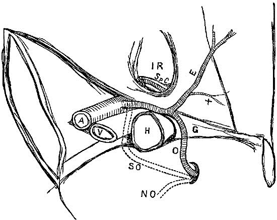
Fig. xxxii. 146
The usual origin of this vessel is from the internal iliac, in which case (Fig. xxxii. n o) it never comes near the sac at all. In certain cases (1 in 3½) it rises from the epigastric, and in a very few (1 in 72) from the external iliac. If rising from either of the two last, it most commonly passes downwards at the outer side of the hernia, in which case (Fig. xxxii. s o) no harm can possibly result; but in a few rare cases, perhaps 1 in every 60 of those operated on, the vessel winds round the hernia (Fig. xxxii. o), crossing at its inner side, and thus may be (and has actually been) divided by a rash incision. With due care, however, and by cutting a very little at a time, even this danger may be avoided.
2. Under what circumstances is it possible or justifiable to reduce a femoral hernia, without previously opening the sac? Only in certain very select cases, where the hernia is recent, the constricting parts lax, the general symptoms very mild, and where there is reason to believe the bowel has completely escaped injury by compression or the taxis. There are both difficulties and dangers in this so-called minor operation:—1. Difficulties, For it is not easy to divide the constriction without the assistance of the finger in the sac, and it is not easy to reduce the contents with the sac unopened, except through a much freer opening than is necessary when the bowel has been fairly exposed. 2. Dangers, Of reducing sac and viscera, together with the strangulation still kept up by tightness in the neck of the sac; or of supposing the sac is emptied while a knuckle of bowel still remains in it, and is strangulated; or, lastly, of reducing the intestine which has already become gangrenous. It is very remarkable how very soon gangrene may come on, in a case of a small recent femoral hernia, in which the fibrous tissues constricting the neck of the sac are tense and undilatable. A protrusion for eight hours has been sufficient to destroy the life of a knuckle of bowel.
A note here on a certain condition very frequent in femoral herniæ, which may occasionally give a good deal of trouble. Symptoms of strangulation have been well marked, yet when the sac is opened nothing is to be seen except a mass of omentum, perhaps tolerably healthy-looking. To reduce this en masse would be very unsafe; it is necessary carefully to unravel it, and disengage the knuckle of bowel which is almost certainly included in it, and which has given rise to the symptoms of strangulation.
Operation for Strangulated Umbilical Hernia.—The operation is practically the same, whether the hernia is a true umbilical one, or one which with more strict accuracy might be called ventral. True umbilical hernia is a disease of infancy and childhood, being almost always congenital, and the viscera protrude through the umbilical aperture. This rarely requires operation, as it may generally be returned with ease, and even cured by a proper bandage and compress. Ventral hernia, commonly called umbilical, is generally a protrusion of viscera through a new preternatural aperture in the fibrous tissues close to the navel, may often attain a large size, is liable to strangulation, and is not easily palliated or cured.
In either case the operation requires a very brief description. If the hernia is small, under the size of a hen's egg, a crucial incision through the thin skin which covers it will thoroughly expose the sac when the flaps are dissected back. The forefinger should then be inserted in the round opening, and the edges cautiously incised in several directions, each incision however being very small.