

 полная версия
A Manual of the Operations of Surgery
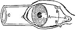
Fig. xi. 86
The original operation of Dieffenbach, now rarely practised, consisted in making an incision, b b, across the tendon, then, by cutting the areolar tissue exposing the insertion of the tendon, and dividing it freely; after which the sclerotic in the neighbourhood was to be cleaned and any band of fibres divided. There are risks on the one hand of a most unseemly exophthalmos with divergent squint, and on the other of a retraction of the semilunar fold, so that the sub-conjunctival operation is always preferable.
Operations for Divergent Squint.—This very serious deformity is often the result of the operation for convergent squint, and is associated with a fixed, leering, and prominent eye, and frequently with most annoying double vision.
1. In a simple case of primary divergent strabismus (very rare) it is sufficient simply to divide the external rectus in the manner already described for division of the internal.
2. If secondary to an operation for convergent squint, the indication is to restore the cut internal rectus to a position on the sclerotic a little behind its previous one, as the cause of the divergence is found in a complete detachment of the internal rectus. This is attempted in various ways.
(1.) Jules Guérin carefully divided the conjunctiva over it, and sought for the remains of the internal rectus, freeing it from its attachments. He then passed a thread through the sclerotic on the outer side of the globe, and by pulling on it and fixing it across the nose, rotated the eye inwards, in the hope that the remains of the internal rectus would secure a new attachment.
(2.) Graefe's modification of this is more certain. Without any minute dissection he merely separated the internal rectus, along with the conjunctiva, and fascia over it, so that it can be pulled forwards, then cut the external rectus, and inverted the eyeball to a sufficient extent by means of a thread passed through the portion of the tendon of the external rectus, which remains attached to the sclerotic. The risk of all these operations, in which both muscles are divided, is protrusion of the eyeball from the removal of muscular tension.
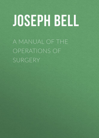
(3.) Solomon's operation for the radical cure of extreme divergent strabismus,87 is at first sight a very curious one. Without going into all the details, the steps are as follows:—
a. A square-shaped flap, with its attached base at the nasal side, is raised, containing the remains of the inner rectus and its adjacent parts.
b. A flap similar in shape and size, but different in the position of its attached base, is made on the other side of the cornea. It is made by dividing the external rectus just behind its tendon, and then reflecting forwards the tendon with its conjunctiva.
c. These two flaps are united over the vertical meridian of the cornea by sutures, three generally being sufficient. This entirely hides the cornea for a time, but eventually shrivels and contracts, and the remnants are to be cut off with scissors three weeks after the operation.
Puncture of the Cornea.—Paracentesis of the Anterior Chamber.—Tapping of the Aqueous Humour.—This very simple operation is in many cases extremely useful. In cases of corneal ulcer, the result either of injury or disease, where there is much pain in the bone, and evidence of tension of the globe, it gives great relief, and when repeated at short intervals greatly hastens a cure. Sperino of Turin recommends its frequent use in cases of chronic glaucoma.
Operation.—The surgeon stands behind the patient, who is seated; the lids being fixed, the upper by the surgeon's left hand, and the lower by an assistant, the cornea is punctured a little in front of the sclerotic margin, either with a broad needle, or, what is as good, a well-worn Beer's knife. Care must be taken on entering the knife, on the one hand, not to wound the iris, which is sometimes arched forwards in the cases of commencing glaucoma, and, on the other, fairly to enter the anterior chamber, not merely split up the layers of the cornea. On withdrawing the cataract knife, the aqueous humour gets out by its side, aided by a slight turn of the knife, sometimes with great force, and in much larger quantity than usual. If the operation has been done by a needle, a blunt probe requires to be introduced on the removal of the needle. Once punctured, the remarkable fact is that the same wound suffices for many succeeding tappings, which are effected by pressing the probe into the wound day after day, sometimes several times a day, with great relief to the symptoms. If the probe is to be used for succeeding evacuations, the operator must be careful to remember the exact spot at which the needle or knife was entered. To facilitate remembering it, it is best, when nothing prevents it, to operate always in the same spot. Sperino chooses the horizontal meridian of the cornea at the temporal side, at the junction of the cornea and sclerotic.
Cataract Operations.—Here we cannot enter into any discussion of the pathology of cataract and the varieties of it. Enough for our purpose to know that the lens is in some cases hard, in others soft, and that thus in the latter it may be removed piecemeal, and by a small incision, while in the former, removal must be almost entire, and by a larger opening.
In cataract, the lens, which should be transparent, has become opaque, and the object of treatment is to get it out of the line of sight, to prevent it from obstructing, now that it can no longer assist sight.
The operations used for this end may be classed under three heads:—
1. Operations for the removal of the lens out of the way without its removal from the eye.—These used to be extensively practised under the name couching, and are of two kinds,—Depression, where the lens is simply pushed down from its place by a needle; Reclination, in which it is shoved backwards (turning on its transverse axis) as well as downwards. These are relics of old surgery, and very rarely practised by any oculists of eminence, as, though easy to perform, and with very flattering immediate results, the risks of chronic inflammation of the whole globe and injury to the retina are very great.
2. For solution.—The Needle Operation.—Suitable (among other cases) especially in congenital cataracts in infants, and in cases of diabetic cataract.
The principle of this operation is that the lens, once the capsule is freely opened in front and the aqueous humour admitted, is found rapidly to become absorbed and disappear, if the cataract has been a soft one.
Operation.—A needle with a lance-shaped head is to be used. It should be so made that the rounded shaft of the needle is just large enough to play freely in the wound made by the broader point, and yet not so small as to allow the aqueous humour to escape rapidly. The pupil has been dilated, the patient is lying on his back, and the globe is fixed by forceps attached to the conjunctiva of the inner side of the eye, and held by an assistant. The surgeon then enters the needle close to the sclerotic margin of the cornea, carries it fairly on in the anterior chamber, till the centre of the pupil is reached. He then, by bringing forward the handle, projects the point backwards against the anterior capsule, which he freely lacerates with the point and edge in several directions.
In infants, where processes of repair go on very rapidly, the whole lens may be freely broken up. In diabetic cataract, or indeed in all cases of solution, where the patient is adolescent or adult, or the eye at all weak, only a small portion of the lens should be attacked at one sitting.
The needle should then be withdrawn gradually and with great care, that the broad axis of the blade be in exactly the same position in which it entered, i.e. flat and parallel with the iris, lest the iris be wounded, entangled, or prolapsed.
The eye is then to be closed for twenty-four hours; if there is much pain, atropia must be freely used.
Varieties in the Operation.—Some use two needles at once for breaking up the lens. Some surgeons prefer to enter the needle through the sclerotic; this complicates the operation and renders it less certain, as the point of the needle is of course out of sight in its progress between the iris and the lens.
Even in children this operation requires in most cases to be repeated at least once, while in adults it may be required at short intervals for many months.
3. By Extraction.—In these operations the lens is at once removed from the eye—
(1.) By linear, or perhaps, more correctly, rectilinear incision. This method is specially suited for cases of soft cataract.
Operation.—A fine spear-shaped needle is very cautiously introduced through the cornea, about a line from its outer margin, and the anterior capsule lacerated, and the lens broken up, great care being taken not to injure the posterior capsule. The pupil must then be kept freely dilated, the wound heals at once, and the aqueous humour reaccumulates.
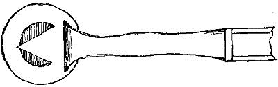
Fig. xii.
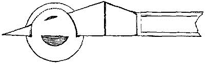
Fig. xiii.
From three to six days after this first operation, a linear incision (Fig. xii.) is made in the outer side of the cornea by a straight stab from a double-edged knife, or rather spear. The size of the incision must vary with the size and consistence of the lens, and can be regulated by the breadth of the knife and the distance to which it is entered. By careful withdrawal of the knife, in many cases a large portion of the soft lens can be removed along with it, and then what remains must be cautiously lifted out by a flat spoon introduced through the wound, and behind the remains of the lens.
Care must be taken lest any of the lens substance remain in the wound; with this precaution the incision generally heals rapidly, and with much less risk of general inflammation of the ball than in the ordinary flap operation of extraction.
Extraction of Soft Cataract by Suction.—Mr. T. P. Teale, of Leeds,88 has invented an instrument by which the removal of soft cataract is made more easy, through a linear incision by suction, applied through the medium of a hollow curette furnished with an india-rubber tube and mouth-piece.
The curette is of the usual size, but is roofed in (instead of being merely grooved) to within one line of its extremity, thus forming a tube flattened above, but terminating in a small cup. This is screwed into an ordinary straight handle, which is hollow for a short distance, far enough to join with a second tube fixed at right angles to the handle, and into which the india-rubber pipe and mouth-piece, through which suction is to be made, is attached. In many cases it seems to serve its purpose extremely well.
Certain points require attention:—1. That the puncture to admit the curette is large enough; 2. That its end be sufficiently rounded; 3. Its open end must be held in the area of the pupil, and not allowed to pass behind the iris, else there is great risk of the iris being drawn in. Among other advantages claimed by its inventor, the chief seems to be a more thorough removal of the lens than by the ordinary means, and consequently less risk of opaque deposit in the posterior capsule.
(2.) Extraction by Flap.—When properly performed in a suitable subject, and when free from accident, this operation is one of the most thoroughly beautiful and satisfactory in the whole domain of surgery; but it is difficult, and liable to many risks which neither skill nor caution can completely guard against.
It is required in many cases of hard cataract, which are amenable neither to solution nor linear extraction.
Operation must be considered in various stages:—
a. To make a flap of cornea large enough to permit of the removal of the entire lens without pressure or bruising. To make it of cornea only, to prevent the escape of the vitreous, and to avoid injury of the iris.
The great difficulty in making the required section of the cornea is, that we are debarred from using scissors or any ordinary knife or scalpel in making it, for this reason, that the sawing movements required in all ordinary cutting are inadmissible here, as any withdrawal of the blade, however slight, would permit evacuation of the aqueous humour, and at once be followed by prolapse of the iris before the knife. Hence we are compelled to make the requisite flap by one steady push of a knife, which, too, must be of such a shape as in its entrance constantly to fill up the wound it makes. Very various shapes and sizes of knives have been proposed, the one called Beer's knife being the sort of model or common parent from which all the others are derived. It is triangular in shape, with a straight back, about 12-10ths of an inch in length, and 4-10ths broad at the base of the blade, tapering at a straight edge from its base to its point, and also diminishing in thickness to the point.
Considerable difference of opinion exists as to the relative merits of an upper or lower section of the cornea. The general view at present seems to be that an upper section is to be preferred; but in cases where the surgeon is not ambidexterous, it is better that he should make the section which lies easiest to his hand than attempt an upper section in a less favourable position.
The patient should be placed flat on his back, the lids should be gently opened, the upper one by the surgeon, the lower one by his assistant, who is to press the lid downwards against the malar bone without exercising any pressure on the ball. The eye should be still further steadied by the conjunctiva and subjacent cellular tissue on the inner side being seized by a pair of catch-forceps, still with no downward pressure on the ball. The point of the knife must then be introduced about a line from the outer sclerotic margin of the transverse diameter of the cornea (Fig. xiii.), the blade being held parallel with the fibres of the iris, pushed steadily across the anterior chamber, and protruded as nearly as possible at the corresponding spot at the inner side of the cornea. The aqueous humour should not escape till the section is completed. If it does, the iris is almost certainly projected forwards and entangled in the blade of the knife, a most annoying accident, and one which is not easily remedied. The books tell us of various manœuvres by pressure or otherwise, by which the iris may be pushed back. Practically, however, if it has once occurred it is not easily saved from being cut. If a small portion only is involved, it is not of much consequence; if a large portion be in danger, it is sometimes necessary to withdraw the knife before the section is completed, and finish it with a probe-pointed, curved bistoury.
If, however, the flap is safely finished, the lids should be gently allowed to close for a few seconds.
On opening them again the surgeon must decide whether the corneal flap is sufficiently large to allow the lens to come out without force; if not, he must enlarge it either by the narrow probe-pointed "secondary knife" or by a pair of sharp scissors. Occasionally the lens, and even a little vitreous humour, may escape at once on the section being completed, but this is not to be desired.
b. Laceration of the Capsule of the Lens.—This is performed by insinuating a sharp curved needle under the corneal flap, avoiding the iris, and then tearing up the anterior capsule through the dilated pupil, the chief point to be attended to being that the capsule be lacerated in its entire length.
c. Removal of the Lens.—This must be done with the most extreme caution and gentleness, lest the vitreous humour be also evacuated. The surgeon's object is to tilt the lens so as to turn it slightly on its transverse axis, and cause the edge nearest the section to rise out of the capsule and appear at the wound. This is best done by gentle pressure at the required spot by the back of the needle, or by a common probe. When the lens begins to protrude the pressure must be very, gentle, lest it be forced out suddenly and the vitreous follow it.
Soft portions of the lens are apt to remain adherent to the wound in the cornea. These must be removed by scoop or probe.
Varieties in the method of Flap Extraction.—Jacobsen of Königsberg in every case gives chloroform. He always makes his flap in the boundary line of the cornea and the sclerotic, through a vascular structure, and he believes that union is on this account more rapid, and after extraction removes that portion of the iris which appears to have been most exposed to bruising during the exit of the lens.
The operation of extraction may in many cases be either preceded or followed by iridectomy, as proposed by Mooren, Von Graefe, and others. The following operation seems to diminish the risks to a very great extent:—
Professor Von Graefe's Operation.—The lids are separated by a speculum, and the eyeball is drawn down by forceps placed immediately below the cornea. The point of a small knife, of which the edge is directed upwards, is inserted at a point fully half a line from the margin of the cornea near its upper part, so as to enter the anterior chamber as peripherally as possible. The point should not be directed at first towards the spot for counterpuncture; nor till the knife has advanced fully three and a half lines within the visible portion of the anterior chamber, should the handle be lowered and the point directed so as to make a symmetrical counterpuncture, which will give the external wound a length of four and a half or five lines. As soon as the resistance to the point is felt to be overcome, showing that the counterpuncture is effected, the knife must at once be turned forward, so that its back is directed almost to the centre of the ideal sphere of the cornea, whether the conjunctiva is transfixed or not, and the scleral border is divided by boldly pushing the knife onwards and again drawing it backwards. This portion of the operation is concluded by the formation of a conjunctival flap a line and a half or two lines in length. A section thus made is almost perpendicular to the cornea, a circumstance much facilitating the passage of the lens, and the line of incision is nearly straight, so that the wound does not gape. The iris should be excised to the very end of the wound, and the capsule most freely opened by a V-shaped laceration. Any lens, even the hardest, may then be removed without the introduction of an instrument into the eye, but Von Graefe's experience shows it to be advisable to assist the evacuation by the hook in about one case in eight. In a certain number of cases the lens will escape without difficulty when the operator presses on the posterior lip of the wound, especially when the back of the spoon is made to glide along the sclera; should this not occur, Von Graefe uses a peculiar blunt hook, or occasionally, though rarely, a spoon. A compressing bandage is applied, and replaced at intervals.89
We are recommended to perform it in two sets of cases:—
1. Those in which the eye is known to be unhealthy and liable to inflammations, specially of iris, retina, or choroid. In cases where the patient has already lost an eye, Von Graefe thinks iridectomy should always precede extraction. In the above, then, it is a precautionary measure, and, if convenient, should be performed three, four, or even six weeks before the extraction.
2. It is recommended to be performed at the same time as extraction in all cases in which the operation has presented any special difficulties, or has not gone smoothly, e.g. in cases where the lens has required much force to expel it, either from the flap of cornea being too small, or from adhesions between the lens and capsule; or, again, in cases in which there is a tendency to prolapse of the iris, in which any of the cortical substance has been necessarily left behind, or in which old adhesions had existed between the iris and capsule, or between the cornea and iris.
Operations for Artificial Pupil.—The cases are by no means unfrequent in which it is necessary to remove or destroy a portion of the iris to admit light to the retina. In cases of excessive prolapse of the iris after extraction of the lens, where the iris has formed adhesions to the wound, and still more frequently in cases where central opacities of the cornea have fairly occluded the natural pupil, the only chance for vision is to enlarge the old one, or make a new pupil by removal of the iris.
Very various operations have been proposed, and exceedingly numerous and complicated instruments invented for this purpose. We can notice here only one or two of the most approved procedures:—
1. Incision is the simplest.
This is practicable and effectual only in cases where the iris is so far healthy as still to retain its contractile power, and so far free from adhesions as to be able to make use of it. The best example of such a case is that of a cataract, in which after extraction a prolapse of the iris has occurred to such an extent as to obliterate the pupil, and where, at the same time, the only adhesions are to the wound, none to the cornea.
Operation.—A double-edged needle is introduced through the cornea near its margin; on arriving at the place where the pupil ought to be, one edge is drawn against the iris, and divides it transversely, if possible, without injuring the lens; the fibres of the iris start back, contract, so that a sufficiently large central pupil may be obtained.
2. Excision.—In the far more frequent cases in which there exist adhesions between iris and cornea, or iris and anterior capsule, incision is not sufficient, and it is necessary to excise a portion of the iris.
The simplest and safest operation is the following:—
The patient recumbent, and the lids held apart by a speculum, the eyeball should be steadied by the forceps of an assistant. A broad cutting needle should then be introduced at the lower or outer edge of the corneal margin. This must be very gently withdrawn so as to retain as much aqueous humour as possible. Into the wound thus made the surgeon must introduce the blunt hook (known as Tyrrell's) at first with its point forwards, then, on arriving opposite the edge of the pupil, which it is intended to enlarge or replace, with its point turned backwards, so as to hook over the edge of the iris and thus drag on it. Once the hook has fairly got hold, it must again be rotated forwards, and withdrawn in the same direction as it was put in. The iris thus pulled out of the wound is to be cut off with a pair of fine scissors, so as to remove a sufficient amount to make a new pupil of the required size.
But in those cases in which the whole or greater part of the pupillary margin is adherent, the blunt hook will not do, because there exists no edge round which to hook it. One of two plans is generally chosen to remedy this:—
(1.) A free incision made with a double-edged needle; through this a pair of canula forceps is introduced, with which a portion of iris is seized and dragged to the external wound; it can then either be cut off or tied (see Iridesis); or,
(2.) A previous attempt may be made to free a portion to form an edge to catch hold of, either by incision or by Corelysis (q.v.)
Iridesis.—Critchett's Operation of Ligature.90—Patient being put under chloroform, the ball is fixed by the wire speculum, and also by a fold of conjunctiva being seized by forceps. An opening is then made with a broad needle through the margin of the cornea, close to the sclerotic, just large enough to admit the canula forceps, with which a small portion of iris close to its ciliary attachment is seized and drawn out; a piece of fine floss silk, previously tied in a small loop round the canula forceps, is slipped down and carefully tightened round the prolapsed portion. This speedily shrinks, and the loop may generally be removed about the second day. The chief advantage claimed for this method is the ease with which the size of the new pupil can be regulated. It is also suitable in cases of conical cornea, where it is wished to change the form of the pupil into a narrow slit.