Полная версия
The Teenage Brain: A neuroscientist’s survival guide to raising adolescents and young adults
So what does being a teenager mean? Man-child, woman-child, quasi-adult? The question is about much more than semantics, philosophy, or even psychology because the repercussions are both serious and practical for parents, educators, and doctors, as well as the criminal justice system, not to mention teens themselves.
Hall, for one, believed adolescence began with the initiation of puberty, and this is why he is considered the founder of the scientific study of adolescence. Although he had no empirical evidence for the connection, Hall knew that understanding the mental, emotional, and physical changes that happen in a child’s transition into adulthood could come only from an understanding of the biological mechanics of puberty.
One of the chief areas of focus in the study of puberty has long been “hormones,” but hormones have gotten a bad rap with parents and educators, who tend to blame them for everything that goes wrong with teenagers. I always thought the expression “raging hormones” made it seem as though these kids had taken a wicked potion or cocktail that made them act with wild disregard for anyone and anything. But we are truly blaming the messenger when we cite hormones as the culprit. Think about it: When your three-year-old has a temper tantrum, do you blame it on raging hormones? Of course not. We know, simply, that three-year-olds haven’t yet figured out how to control themselves.
In some ways, that’s true of teenagers as well. And when it comes to hormones, the most important thing to remember is that the teenage brain is “seeing” these hormones for the first time. Because of that, the brain hasn’t yet figured out how to modulate the body’s response to this new influx of chemicals. It’s a bit like taking that first (and hopefully last!) drag on a cigarette. When you inhale, your face flushes; you feel light-headed and maybe even a bit sick to your stomach.
Scientists now know that the main sex hormones—testosterone, estrogen, and progesterone—trigger physical changes in adolescents such as a deepening of the voice and the growth of facial hair in boys and the development of breasts and the beginning of menstruation in girls. These sex hormones are present in both sexes throughout childhood. With the onset of puberty, however, the concentrations of these chemicals change dramatically. In girls, estrogen and progesterone will fluctuate with the menstrual cycle. Because both hormones are linked to chemicals in the brain that control mood, a happy, laughing fourteen-year-old can have an emotional meltdown in the time it takes her to close her bedroom door. With boys, testosterone finds particularly friendly receptors in the amygdala, the structure in the brain that controls the fight-or-flight response—that is, aggression or fear. Before leaving adolescence behind, a boy can have thirty times as much testosterone in his body as he had before puberty began.
Sex hormones are particularly active in the limbic system, which is the emotional center of the brain. That explains in part why adolescents not only are emotionally volatile but may even seek out emotionally charged experiences—everything from a book that makes her sob to a roller coaster that makes him scream. This double whammy—a jacked-up, stimulus-seeking brain not yet fully capable of making mature decisions—hits teens pretty hard, and the consequences to them, and their families, can sometimes be catastrophic.
While scientists have long known how hormones work, only in the past five years have they been able to figure out why they work the way they do. Because sex hormones are present at birth, they essentially hibernate for more than a decade. What, then, triggers them to begin puberty? A few years ago, researchers discovered that puberty is initiated by what appears to be a game of hormonal dominoes, which begins with a gene producing a single protein, named kisspeptin, in the hypothalamus, the part of the brain that regulates metabolism. When that protein connects with, or “kisses,” receptors on another gene, it eventually triggers the pituitary gland to release its storage of hormones. Those surges of testosterone, estrogen, and progesterone in turn activate the testes and ovaries.
After sex hormones were discovered, for the rest of the twentieth century they became the dominant theory of, and favorite explanation for, adolescent behavior. The problem with this theory is that teenagers don’t have higher hormone levels than young adults—they just react differently to hormones. For instance, adolescence is a time of increased response to stress, which may in part be why anxiety disorders, including panic disorder, typically arise during puberty. Teens simply don’t have the same tolerance for stress that we see in adults. Teens are much more likely to exhibit stress-induced illnesses and physical problems, such as colds, headaches, and upset stomachs. There is also an epidemic of symptoms ranging from nail biting to eating disorders that are commonplace in today’s teens. We have a tsunami of input coming at teens from home, school, peers, and, last but not least, the media and Internet that is unprecedented in the history of mankind. Why are adults less susceptible to the effect of all this stimulation? In 2007, researchers at the State University of New York (SUNY) Downstate Medical Center reported that the hormone tetrahydropregnanolone (THP), usually released in response to stress to modulate anxiety, has a reverse effect in adolescents, raising anxiety instead of tamping it down. In an adult, this stress hormone acts like a tranquilizer in the brain and produces a calming effect about a half hour after the anxiety-producing event. In adolescent mice, THP is ineffective in inhibiting anxiety. So anxiety begets anxiety even more so in teens. There is real biology behind that.
In order to truly understand why teenagers are moody, impulsive, and bored; why they act out, talk back, and don’t pay attention; why drugs and alcohol are so dangerous for them; and why they make poor decisions about drinking, driving, sex—you name it—we have to look at their brain circuits for answers. The elevated secretion of sex hormones is the biological marker of puberty, the physiological transformation of a child into a sexually mature human being, though not yet a true “adult.”
While hormones can explain some of what is going on, there is much more at play in the teenage brain, where new connections between brain areas are being built and many chemicals, especially neurotransmitters, the brain’s “messengers,” are in flux. This is why adolescence is a time of true wonder. Because of the flexibility and growth of the brain, adolescents have a window of opportunity with an increased capacity for remarkable accomplishments. But flexibility, growth, and exuberance are a double-edged sword because an “open” and excitable brain also can be adversely affected by stress, drugs, chemical substances, and any number of changes in the environment. And because of an adolescent’s often overactive brain, those influences can result in problems dramatically more serious than they are for adults.
2
Building a Brain
The human body is amazing, the way it neatly tucks all these complex organs into this finite space and connects them into one smoothly functioning system. Even the average human brain is said by many scientists to be the most complex object in the universe. A baby brain is not just a small adult brain, and brain growth, unlike the growth of most other organs in the body, is not simply a process of getting larger. The brain changes as it grows, going through special stages that take advantage of the childhood years and the protection of the family, then, toward the end of the teen years, the surge toward independence. Childhood and teen brains are “impressionable,” and for good reason, too. Just as baby chicks can imprint on the mother hen, human children and teens can “imprint” on experiences they have, and these can influence what they choose to do as adults.
Such was the case with me. I “imprinted” on neuroscience and medicine pretty early on. My experiences cultivated in me a curiosity that I found irresistible, sustaining me from my high school years through medical school and graduate research, and to this very day. I grew up the oldest of three children in a comfortable family home in Connecticut, just forty minutes from Manhattan. I happened to live in Greenwich, which even back then was the home of actors, authors, musicians, politicians, bankers, and the independently wealthy. The actress Glenn Close was born there, President George H. W. Bush grew up there, and the great bandleader Tommy Dorsey died there.
My parents were from England; they had immigrated after World War II, and my dad came over after medical school in London to do his urological surgery residency at Columbia. To them, Greenwich seemed a great place to settle within commuting distance of New York City. It was a matter of convenience, and they were pretty oblivious to the celebrity status of the town. Perhaps because of my father, I was open-minded about learning math and science. For me a major “imprinting” moment that propelled me in the direction of medicine was a ninth-grade biology class at Greenwich Academy. The best part to me, memorable in fact, was when we each got a fetal pig to dissect. While many of my classmates slumped in their seats at the proposition of slicing up these small mammals, some rushing to the girls’ washrooms with waves of nausea, a few of us jumped into the task at hand. It was one of those defining moments. The scientists had separated from those destined to be the writers, lawyers, and businesspeople of the future.
Injected with latex, the pigs’ veins and arteries visibly popped out with their colorful hues of blue and red. I’m a very visual person; I also like thinking in three dimensions. That visual-spatial ability comes in handy with neurology and neuroscience. The brain is a three-dimensional structure with connections between brain areas going in every direction. It helps to be able to mentally map these connections when one is trying to determine where a stroke or brain injury is located in a patient presenting with a combination of neurological problems—definitely a plus for a neurologist. Actually, that’s how the minds of most neurologists and neuroscientists work. We’re a breed that tends to love to look for patterns in things. I’ve never met a jigsaw puzzle, in fact, that I didn’t like. My attraction to neuroscience in high school and college began at a time before CT scans and MRIs, when a doctor had to imagine where the problem was inside the brain of a patient by picturing the organ three-dimensionally. I’m good at that. I like being a neurological detective, and as far as I’m concerned, neuroscience and neurology turned out to be the perfect profession for me to make use of those visuospatial skills.
If the human brain is very much a puzzle, then the teenage brain is a puzzle awaiting completion. Being able to see where those brain pieces fit is part of my job as a neurologist, and I decided to apply this to a better understanding of the teen brain. That’s also why I’m writing this book: to help you understand not only what the teen brain is but also what it is not, and what it is still in the process of becoming. Among all the organs of the human body, the brain is the most incomplete structure at birth, just about 40 percent the size it will be in adulthood. Size is not the only thing that changes; all the internal wiring changes during development. Brain growth, it turns out, takes a lot of time.
And yet the brain of an adolescent is nothing short of a paradox. It has an overabundance of gray matter (the neurons that form the basic building blocks of the brain) and an undersupply of white matter (the connective wiring that helps information flow efficiently from one part of the brain to the other)—which is why the teenage brain is almost like a brand-new Ferrari: it’s primed and pumped, but it hasn’t been road tested yet. In other words, it’s all revved up but doesn’t quite know where to go. This paradox has led to a kind of cultural mixed message. We assume when someone looks like an adult that he or she must be one mentally as well. Adolescent boys shave and teenage girls can get pregnant, and yet neurologically neither one has a brain ready for prime time: the adult world.
The brain was essentially built by nature from the ground up: from the cellar to the attic, from back to front. Remarkably, the brain also wires itself starting in the back with the structures that mediate our interaction with the environment and regulate our sensory processes—vision, hearing, balance, touch, and sense of space. These mediating brain structures include the cerebellum, which aids balance and coordination; the thalamus, which is the relay station for sensory signals; and the hypothalamus, a central command center for the maintenance of body functions, including hunger, thirst, sex, and aggression.
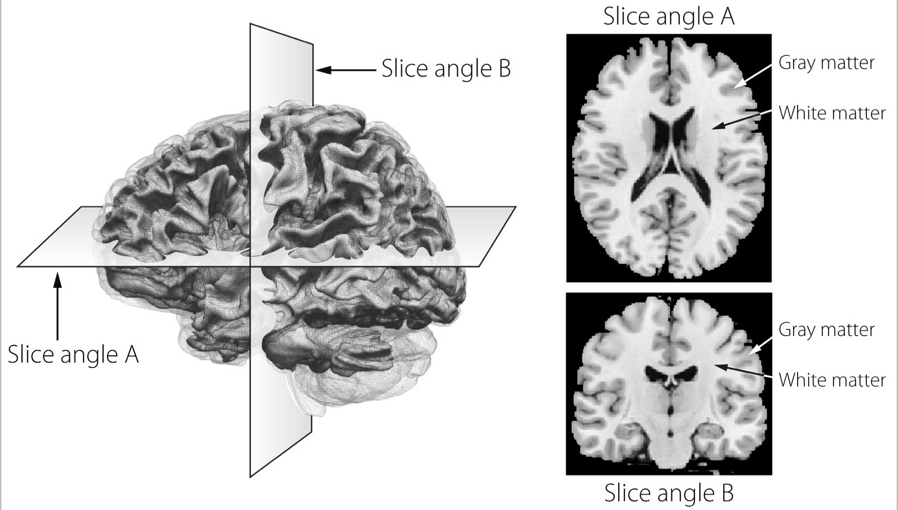
I have to admit that the brain is not very exciting to look at. Sitting atop the spinal cord, it is light gray in color (hence the term “gray matter”) and has a consistency somewhere between overcooked pasta and Jell-O. At three pounds, this wet, wrinkled tissue is about the size of two fists held next to each other and weighs no more than a large acorn squash. The “gray matter” houses most of the principal brain cells, called neurons: these are the cells responsible for thought, perception, motion, and control of bodily functions. These cells also need to connect to one another, as well as to the spinal cord, for the brain to control our bodies, behavior, thoughts, and emotions. Neurons send most of their connections to other neurons through the “white matter” in the brain. The commonly used brain imaging tool, magnetic resonance imaging, or MRI, shows the distinction between gray and white matter beautifully. On the outside surface, the brain has a rippled structure. The valleys or creases are referred to as sulci and the hills are referred to as gyri. Figure 1 shows an image from a brain’s MRI scan, like those done on patients. There are two sides to the brain, each called a hemisphere. (When an MRI image shows a cut across the middle in one direction or the other [slice angles A and B], it is easier to see the two sides.) The most superficial layer of the brain is called the cortex and it is made up of the gray matter closest to the surface, with the white matter located beneath it. The gray matter is where most of the brain cells (neurons) are located. The neurons connect directly to those close by, but in order to connect to neurons in other parts of the brain, in the other hemisphere, or in the spinal cord to activate muscles and nerves in our face or body, the neurons send processes down through the white matter. The white matter is called “white” because in real life and also in the MRI scans its color is light, owing to the fact that the neuron processes running through here are coated with a fatty insulator-like substance called myelin, which truly is white in color.
As I said before, sheer size—or even weight, for that matter—doesn’t mean everything. A whale brain weighs about twenty-two pounds; an elephant brain about eleven. If intellect were determined by the ratio of brain weight to body weight, we’d be losers. Dwarf monkeys have one gram of brain matter for every twenty-seven grams of body matter, and yet the ratio for humans is one gram of brain weight to forty-four grams of body weight. So we actually have less brain per gram of body weight than some of our primate cousins. It is the complexity of the way neurons are hooked up to one another that matters. Another example of how little the weight of the brain has to do with its functioning, at least in terms of intelligence, is that the human female brain is physically smaller in size than the male brain but IQ ranges are the same for the two sexes. At only 2.71 pounds, the brain of Albert Einstein, indisputably one of the greatest thinkers of the twentieth century, was slightly underweight. But recent studies also show that Einstein had more connections per gram of brain matter than the average person.

FIGURE 1. The Basics of Brain Structure: A magnetic resonance imaging (MRI) scan of a brain. The horizontal and vertical cross sections (slice angles A and B) show the cortex (gray matter) on the surface and the white matter underneath.
The size of the human brain does have a lot to do with the size of the human skull. Basically, the brain has to fit inside the skull. As a neurologist, you have to measure the size of children’s heads as they grow up. I have to admit there were occasions when I did this with my own sons—just like noting changes in their height—to make sure they were on track and in the normal range for skull size. When they were older, they thought I was nuts, of course, but when they were babies and toddlers, I just couldn’t resist coming at them with a tape measure I’d take from my sewing kit, then trying to get them to stop squiggling free so I could take just one more measurement. The truth is, skull size doesn’t tell us a lot. It’s a gross measurement, and the skull can be large or small for a variety of reasons. There are disorders in which the head is too big and disorders in which the head is too small. The most important characteristic of the skull is that it limits the size of the brain. Eight of the twenty-two bones in the human skull are cranial, and their chief job is to protect the brain. At birth, these cranial bones are only loosely held together with connective tissue so that the head can compress a bit as the baby moves through the birth canal. The skull bones are loosely attached and have spaces between them: one of these is the “soft spot” all babies have at birth, which closes during the first year of life as the bones fuse together. Most growth in head size occurs from birth to seven years, with the largest increase in cranial size occurring during the first year of life because of massive early brain development.
So with a fixed skull size, human evolution did its best to jam as much brain matter inside as possible. Homo erectus, from whom the modern human species evolved, appeared about two million years ago. Its brain size was only about 800 to 900 cubic centimeters, as opposed to the approximately 1,500 cubic centimeters of today’s Homo sapiens. With modern human brains nearly double the size of these ancestors’, the skull had to grow as well and, in turn, the female pelvis had to widen to accommodate the larger head. Evolution accomplished all of this within just two million years. Perhaps that’s why the brain’s design, while extraordinarily ingenious, also gives a bit of the impression that it was updated on the fly. How else to explain the cramped conditions? Like too many clothes crammed in too small a closet, the evolution-sculpted brain looks like a ribbon repeatedly folded and pressed together. These folds, with their ridges (gyri) and valleys (sulci), as seen in Figure 1, give the human brain an irregular surface appearance, the result of all that tight packing inside the skull. Not surprisingly, humans have the most complex brain folding structure of all species. As you move down the phylogenic scale to simpler mammals, the folds begin to disappear. Cats and dogs have some, but not nearly as many as humans do, and rats and mice have virtually none. The smoother the surface, the simpler the brain.
While the brain looks fairly symmetrical from the outside, inside there are important side-to-side differences. No one is really sure why, but the right side of your brain controls the left side of your body and vice versa; this means that the right cortex governs the movements of your left eye, left arm, and left leg and the left cortex governs the movements of your right eye, right arm, and right leg. For vision, the input from the left side of the visual field goes through the right thalamus to the right occipital cortex, and information from the right visual field goes to the left. In general, visual and spatial perception is thought to be more on the right side of the brain.
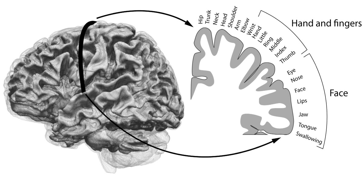
The image of the body, in fact, can actually be “mapped” onto the surface of the brain, and this map has been termed the “homunculus” (Latin for “little man”). In the motor and sensory cortex, the different areas of the body get more or less real estate depending upon their functional importance. The face, lips, tongue, and fingertips get the largest amount of space, as the sensation and control necessary for these areas have to be more accurate than for other areas such as the middle of the back.
An early-twentieth-century Canadian neuroscientist, Wilder Penfield, was the first to describe the cortical map, or homunculus, which he did after doing surgery to remove parts of the brain that caused epileptic seizures. He would stimulate areas of the surface to determine which parts would be safe to remove. Stimulating one area would cause a limb or facial part, for instance, to twitch, and having done this on many patients he was able to create a standard map.

FIGURE 2. The “Homunculus”: A “map” of the brain illustrating the regions that control the different body parts.
The amount of brain area devoted to a given body part varies depending on how complicated its function is. For instance, the area given to hands and fingers, lips and mouth, is about ten times larger than that for the whole surface of the back. (But then, what do you do with your back anyway—except bend it?) This way all the brain regions for the same part of the body end up in close proximity to one another.
My undergraduate thesis at Smith College in Northampton, Massachusetts, examined several of those areas of the brain given over to individual body parts and whether overstimulation of one of the body’s limbs might result in more brain area devoted to that part. This was actually an early experiment in brain plasticity, to see if the brain changed in response to outward stimulation. Many impressive studies that have been done since the late 1970s back up the whole concept of imprinting. Some of the most famous work, which inspired me to do my little undergraduate thesis, was done by a pair of Harvard scientists named David Hubel and Torsten Wiesel. The term that started to be used was “plasticity,” meaning that the brain could be changed by experience—it was moldable, like plastic. Hubel and Wiesel showed that if baby kittens were reared with a patch on one eye during the equivalent of their childhood years (they looked sort of like pirate kittens!), for the rest of their lives they were unable to see out of the eye that had been patched. The scientists also saw that the brain area devoted to the patched eye had been partially taken over by the open eye’s connections. They did another set of experiments where the kittens were raised in visual environments with vertical lines and found that their brains would respond only to vertical lines when they were adults. The point is that the types of cues and stimuli that are present during brain development really change the way the brain works later in life. So my experiment in college showed basically the same thing, not for vision, but rather for touch.
I actually had some fun showing off this cool imprinting effect in our everyday lives. Our beloved cat had died at the ripe old age of nineteen, and we all missed her so. Of course, it didn’t take long before Andrew, Will, and I were at the local animal shelter, looking at kittens to bring home. We fell in love with the runt of a litter and brought home the most petite and needy little tabby kitten you could ever imagine. The boys came up with a name: Jill. Jill was always on our laps; she was a very people-friendly cat. I remembered the experiments on brain plasticity and said to Andrew and Will, when we hold her, let’s massage her paws and see if she becomes a more coordinated cat. So anytime we had her in our laps, we would massage her paws with our hands, spreading them out, touching the little “fingers” that cats’ paws contain. Sure enough, Jill started to use her paws much more than any other cat we had ever had (and I have had back-to-back cats since the age of eight). She used her paws for things most cats don’t. She was very “paw-centric,” going around the house batting small objects off tables and taking obvious pleasure in watching them hit the ground. This was a source of consternation as not all the things she knocked off were unbreakable. She also often used her left paw to eat, gingerly reaching into the cat food can with her paw and scooping up food to bring to her mouth. Watching her, we started to notice that she almost always used her left paw to do these things. We had a left-pawed cat! Then suddenly we realized that when we picked her up to massage her paws, she was facing us and because we are all right-handed, we were always stimulating her left paw much more than her right! Home neuronal plasticity demonstration project accomplished. I know if we could have looked into her brain, we’d have seen that she had more brain space given over to her paws, and especially her left paw, than the average cat. This same phenomenon of reallocating brain space based on experience during life happens in people, too. We call this part of life the critical period, when “nurture,” that is, the environment, can modify “nature.” But more on that later.


