

Полная версия
Myocardial torsion
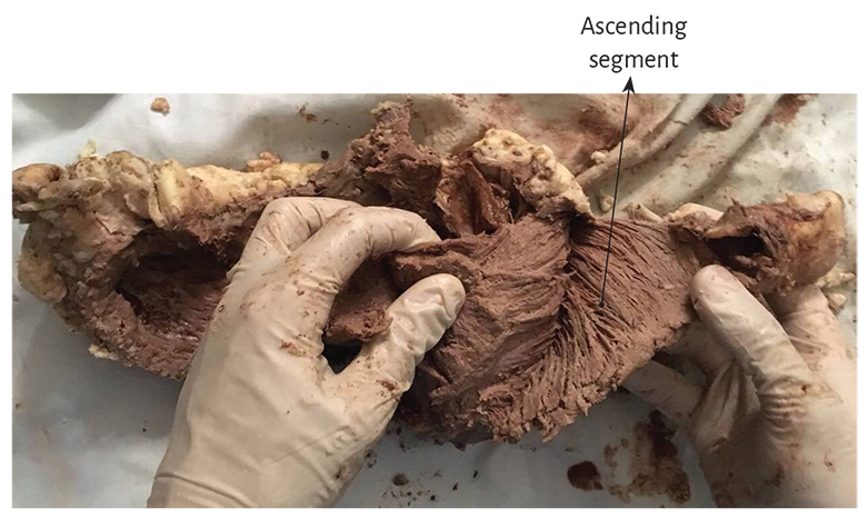
Figure 1.18. The figure shows the ascending segment, which is going to insert in the cardiac fulcrum (bovine heart) (see Figure 1.19).
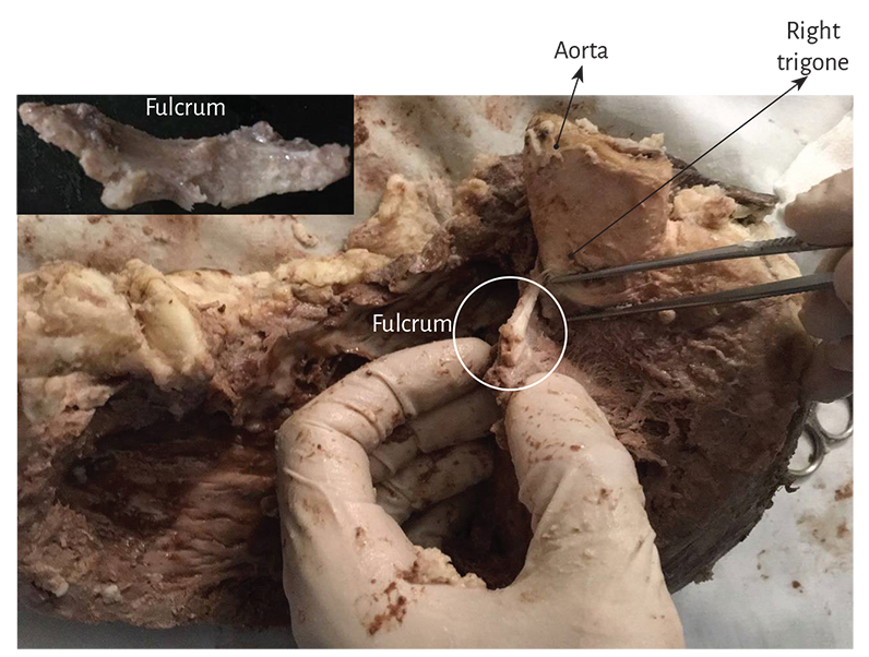
Figure 1.19. Cardiac fulcrum below the right trigone (bovine heart). The insert shows the resected piece.
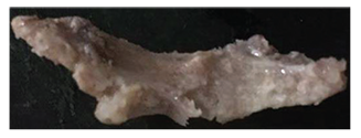
Figure 1.20. Resected cardiac fulcrum
(bovine heart).
The findings in the human hearts are surprising from the point of view of interpretation, since it is logic to consider its presence in all the evolutionary mammalian chain. It should be assumed that this structure, analyzed in different species, has the common function of providing support to the myocardial band to generate the power needed by any muscle, which is different in diverse mammals. Therefore, its presence is constant in all the hearts studied, both in bovids and humans, but its structural characteristic is different, and this diversity in the intimal analysis of the cardiac fulcrum is undoubtedly associated with the resistance it must oppose to the activity of the myocardial band in hearts of different sizes.
The analysis of a 10-year-old human heart showed in the same place a myxoid-cartilaginous formation with approximately 2 cm diameter (Figure 1.21). A similar finding, both in structure and location, occurred in the heart of a 23-week gestation human fetus (Figures 1.22 and 1.23).
A fact defying logic is having found in the adult human heart a formation presenting consistent characteristics, both to observation and palpation, in the same location and with similar triangular morphology (Figures 1.24 to 1.27). However, the histological analysis revealed a matrix similar to that of a tendon.
The macroscopic and microscopic observation reveal the muscle fiber attachment to this solid, homogeneous nucleus, which is closely related with the aortic wall on the side of the tricuspid valve. Its configuration has been confirmed histologically. We have called his structure, origin and end of the myocardial band, cardiac fulcrum, as a parallelism and tribute to the definition of the supporting point of a lever expressed by Archimedes of Syracuse (Greek, 288 B.C. – 212 B.C.). It is located anterior to the central fibrous ridge (right trigone) and it clearly shows that the myocardial fibers of the right segment originate in its structure (Figure 1.16) same as the ascending segment courses to meet it in order to attach (Figures 1.18 and 1.19). It should be noted that to visualize the cardiac fulcrum it is essential to unfold the myocardial band. This osseous, cartilaginous or tendinous structure was always present and with the same morphology in all the hearts analyzed by us, albeit with different histological texture. No description of its characteristics or function has been reported in the literature, except the mere mention of its presence as os cordis in bovids.
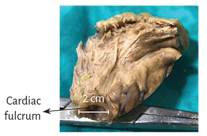
Figure 1.21. Cardiac fulcrum in a 10-year-old human heart (explant).
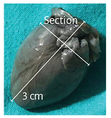
Figure 1.22. Human embryo heart (23-week gestation) showing sectioning planes.
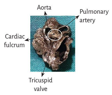
Figure 1.23. Cardiac fulcrum in a 23-week gestation human embryo hear.
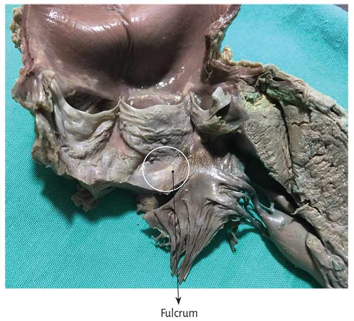
Figure 1.24. Fulcrum in the adult human heart.
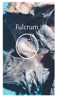
Figure 1.25. Fulcrum in
the adult human heart.
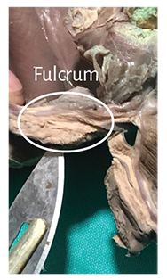
Figure 1.26. Fulcrum resected from
an adult human heart.
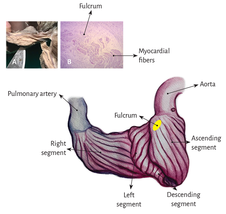
Figure 1.27. The drawing shows the myocardial band arrangement at the beginning of its unfolding. The pulmonary artery and the right segment have been separated from the fulcrum to show its intermediate location between the right segment (anterior location) and the ascending segment (posterior location). A: macroscopic image of the fulcrum in the adult human heart. B: microscopic image of the human fulcrum. Notice the myocardial fibers inserting into the tendinous fulcrum matrix
Histology. The microscopic analysis of the bovine cardiac fulcrum shows a trabecular osteochondral matrix with segmental lines. Its general structure resembles the metaphyseal growth of the long bones (Figures 1.28 and 1.29) and increased magnification reveals bone trabeculae with osteoblasts and segmental lines secondary to bone apposition (Figure 1.30).
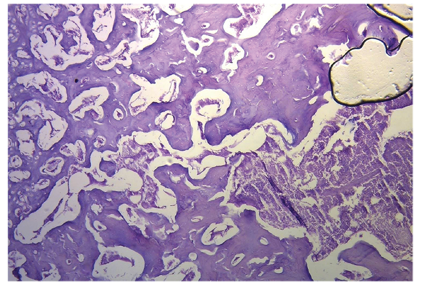
Figure 1.28. Histological section of the cardiac fulcrum showing trabecular bone tissue with osteologic segmental lines (bovine heart). Hematoxylin-eosin stain.
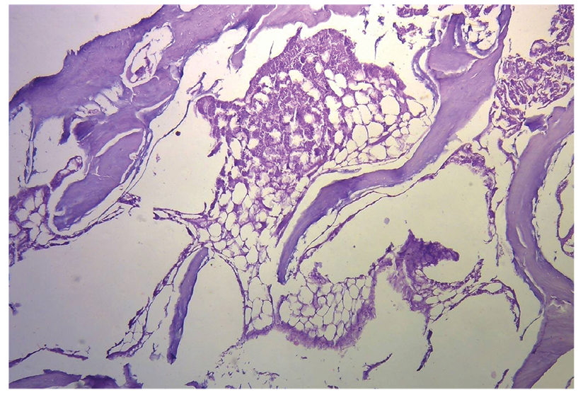
Figure 1.29. Mature trabecular bone forming the cardiac fulcrum tissue (bovine heart). Hematoxylin-eosin stain at low magnification (10×).
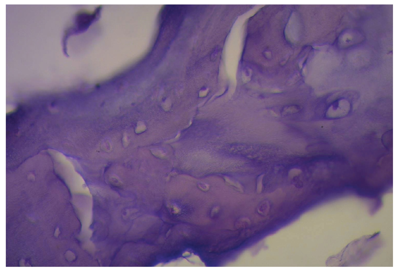
Figure 1.30. Cardiac fulcrum trabecular bone with osteoblasts and segmental lines. The structure constitutes the trabecular bone tissue scaffolding similar to the metaphyseal area of growth in long bones. The histological section also shows bone trabeculae with osteoblasts and segmental lines secondary to bone apposition (bovine heart). Hematoxylin-eosin stain at high magnification (40×).
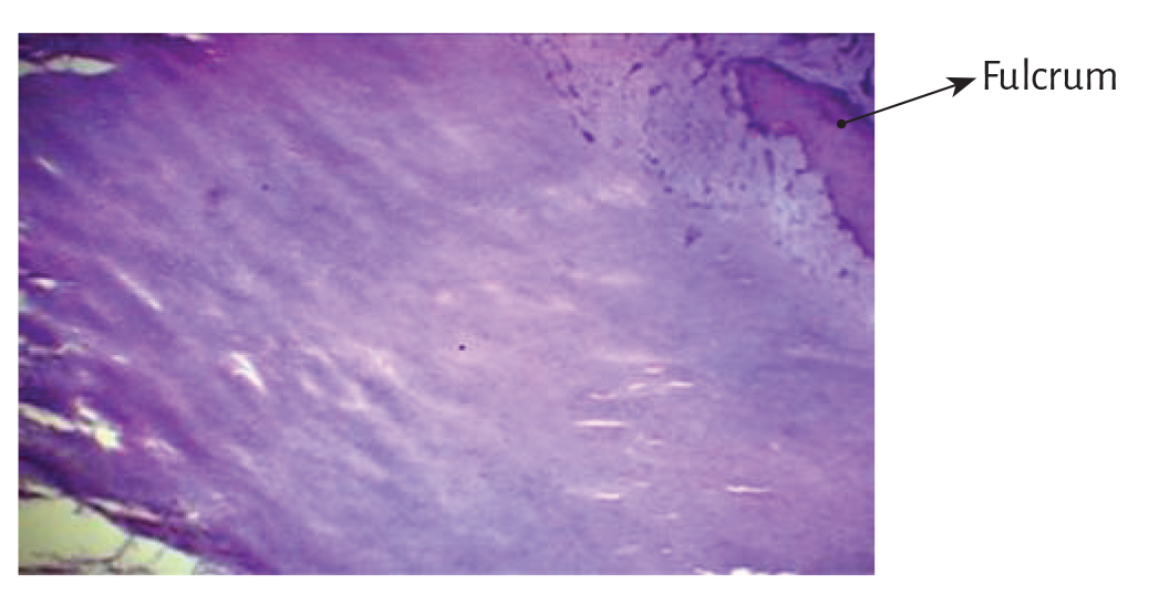
Figure 1.31. Ten year old human heart. Central area of the fulcrum formed by chondroid tissue in a 10-year-old human heart. Hematoxylin-eosin stain.
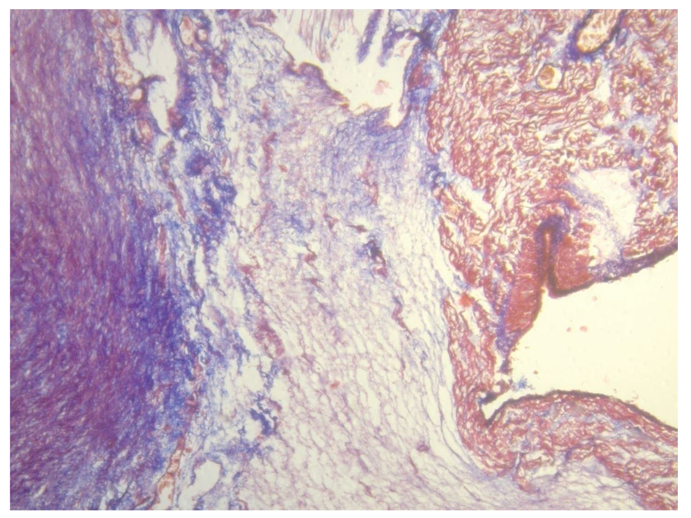
Figure 1.32. Cardiac fulcrum in a 23-week gestation fetus showing prechondroid bluish areas in a myxoid stroma. Masson’s trichrome staining technique.
In the 10-year-old human heart, the histological findings in the cardiac fulcrum were associated with that early age. Figure 1.31 shows a central zone of the fulcrum formed by chondroid tissue. Given the 10-year-old age, it is logical that the fulcrum is smaller and characterized by more chondroid than bone tissue. This finding was repeated in a 23-week old human fetus with the characteristic prechondroid bluish areas in a myxoid stroma (Figure 1.32).
The osseous structure in the bovine os cordis and its relationship with the myxoid-chondroid cardiac fulcrum texture in human hearts, even in gestational stages, is rational for the interpretation analysis. This disparity is associated with the different age evolution from chondroid to osseous material and by the greater force developed in bovids requiring a more rigid supporting point.
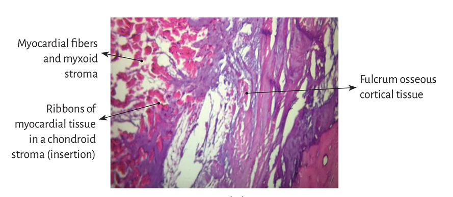
Figure 1.33. Myocardial insertion in
the cardiac fulcrum. Bovine heart.
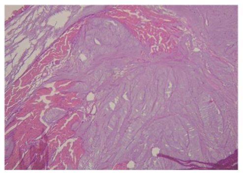
Figure 1.34. Insertion of myocardial fibers in the fulcrum
chondroid tissue of a bovine heart.
However, the histological analysis of the fulcrum in adult human hearts evidenced a tendinous collagenous matrix, needing an additional clarification. In principle, there is constancy in the detection, site and morphology of the fulcrum in all the hearts analyzed. This means that from a functional point of view, its presence is akin to myocardial band insertion, as established in the histological analysis, becoming a solid point of interpretation to achieve its biomechanical function. And we find this demonstration when the histological examination is directed to the myocardial insertion in the osseous, chondroid or tendinous fulcrum. All the hearts studied presented this myocardial attachment to the rigid structure of the fulcrum, integrating a cardiomyocyte-matrix unit, regardless of its osseous, cartilaginous or tendinous nature (Figures 1.33 to 1.38).
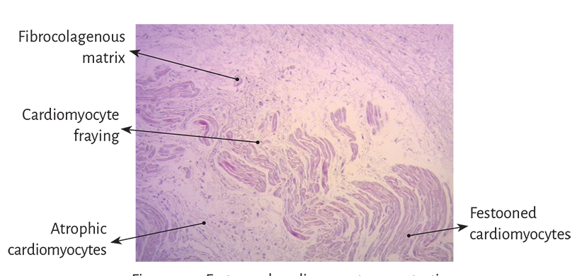
Figure 1.35. Festooned cardiomyocytes penetrating the fibro-colagenous matrix (adult human heart).
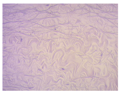
Figure 1.36. Fibrocolagenous matrix at higher magnification (adult human heart).
At this point, fundamental questions arise. Why does the human fulcrum have characteristics similar to a tendon, despite it fulfills the same function of attaching the myocardial band? Why does it not have the same structure of the fetal or child human heart?
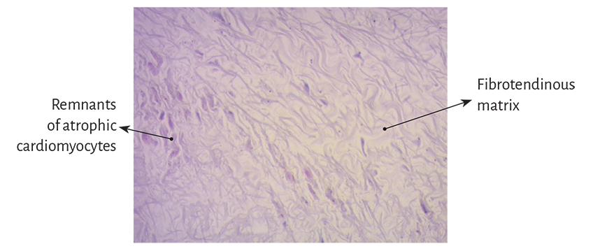
Figure 1.37. Festooned colagenous fibers integrating the fibrotendinous matrix of the fulcrum (adult human heart).
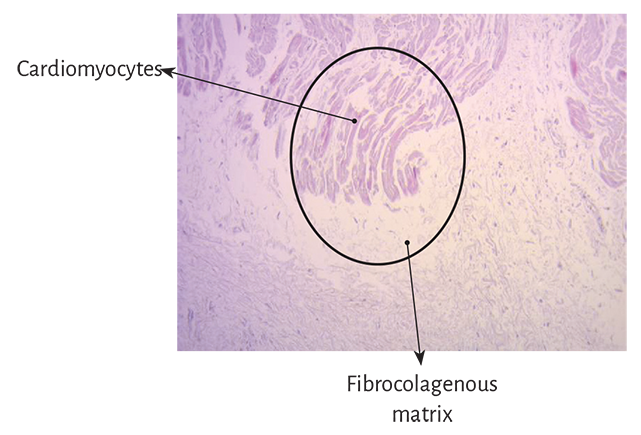
Figure 1.38. Cardiomyocytes penetrating the fibrocolagenous tissue (adult human heart). The circle details the insertion site.
Our interpretation is that perhaps the osseous fulcrum is a vestigial organ specific of mammalian evolution. A vestigial structure must be understood as the preservation during the evolutionary process of genetically established attributes which have lost all or part of their ancestral function in a certain species. (45) As a result, it is found in the initial process of human gestation, but later loses its osseous character, remaining as a tendinous matrix able to achieve myocardial band insertion to attain a muscle power which is much lower than that of larger mammals. Recall that in bovids the fulcrum found in this investigation is of bone nature (Figures 1.28 to 1.30).

Figure 1.39. Collagen tissue and elastoid interstitial tissue are observed. There is no inclusion of cardiomyocytes.
A histological analysis has also been carried out on the trigones trying to find cardiomyocytes in them, as a possibility of insertion of the band in these structures. In our investigation, only collagen tissue was observed without cardiomyocytes in the trigones, confirming that the fulcrum is the support of the band, both in its beginning and in its termination (Figure 1.39).
Fulcrum imaging studies. Bovine hearts studied with computed tomography (Figures 1.40 to 1.42), magnetic resonance imaging (Figures 1.43 and 1.44) and X-rays (Figure 1.45) identified the osteochondral nucleus found in dissection, with the same morphology and analogous size.
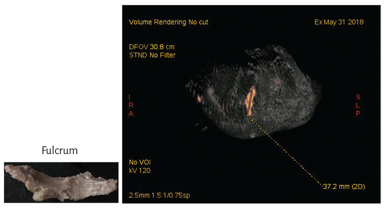
Figure 1.40. Computed tomography. A hyperdense, approximately 3.7 cm long image, with 298 HU density is seen in the interventricular septum adjacent to the root of the aorta (bovine heart).
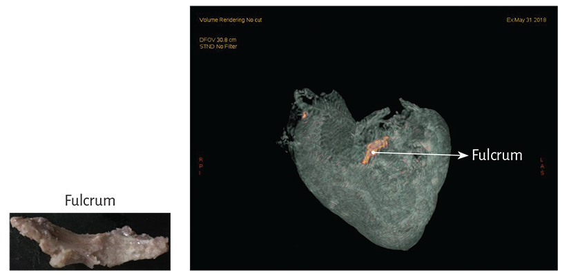
Figure 1.41. Computed tomography of the cardiac fulcrum (bovine heart). The inset shows the resected fulcrum.
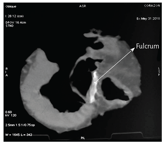
Figure 1.42. Computed Tomography. In the area indicated by the arrow there is an image adjacent to the aortic root on the interventricular septum (bovine heart).
Interpretation. The anterior portion of the cardiac fulcrum is associated with the pulmo-tricuspid cord (Figures 1.15 to 1.17). It is situated between the pulmonary artery and the tricuspid valve and in front of the aortic valve at the level of the right coronary artery, giving rise to the origin of the myocardial band (right segment). Since the right segment separates into two main groups of fibers forming the paraepicardial and paraendocardial bundles, a raphe is defined between then called the pulmo-tricuspid cord.
According to histological studies, analyzed in our investigations, the osseous, chondroid or tendinous fulcrum is close to the tricuspid valve (right), the aorta (posterior) and the pulmo-tricuspid cord (anterior). In order to find it, it is necessary to unfold the myocardial band stripping the aortic root (Figure 1.27) and separating the muscle plane of the descending segment from that of the ascending segment. Then, the latter must be followed up to its insertion in the fulcrum. Before affixing to it, the ascending segment gathers into a bundle whose most external fibers form a curve to attain this attachment, while the most internal fibers enter directly without describing any deviation.
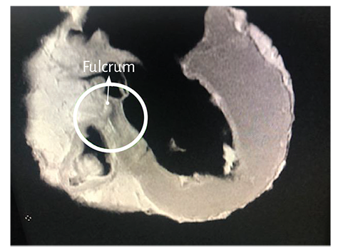
Figure 1.43. Nuclear magnetic resonance image in the bovine heart.
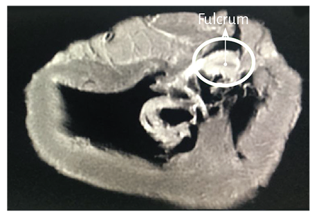
Figure 1.44. Nuclear magnetic resonance image in the bovine heart.
The pulmo-tricuspid cord is located in front of an area that surrounds the anterior two-thirds of the aortic annulus circumference in a U shape, and its open end (posterior) is occupied by the anterior cusp of the mitral valve. This tissue has a trigone at each end. The right fibrous trigone (central fibrous mass) is the most prominent and is placed between the tricuspid (right) and aortic (posterior) orifices and the pulmo-tricuspid cord (anterior) (Figure 1.9). The left less prominent trigone is located between the mitral valve orifice (left) and the aorta. There is no connective tissue in the continuity of the aortic orifice with the posterior cusp of the mitral valve, as laterally, the two fibrous masses extend by means of a band of connective tissue that partially surrounds the mitral valve orifice to progressively vanish. The septal cusp of the mitral valve is found as a wedge between both trigones and can be considered an extension of the atrial endocardium.
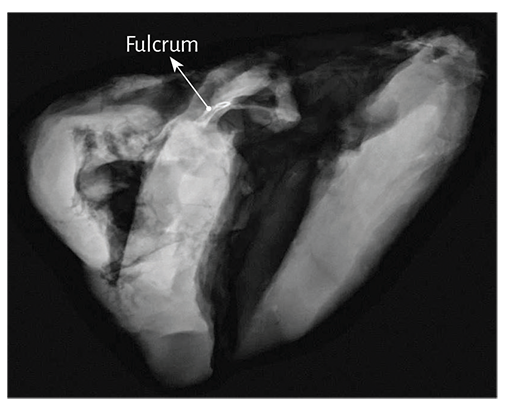
Figure 1.45. Radiologic image of the cardiac fulcrum with mammography technique in the bovine heart.
The cardiac fulcrum is adjacent and anteroinferior to the right trigone, constituting a solid, homogeneous structure to palpation with osseous, chondroid or tendinous histology. It is the site where the right segment and the ascending segment fibers attach, origin and end of the myocardial band. These insertions must be understood as the supporting point of the myocardial band to fulfill its hemodynamic function.
Regarding the study on this point of attachment which we have termed cardiac fulcrum, it must be considered as an organic structure that supports the band, allowing it to develop the necessary force for the fundamental rotational motions of the left ventricle. (131, 132) We should not forget that the heart pendulates within the thoracic cavity and its points of attachment are the origin and end of the myocardial muscle band, very close to each other at the root of the aorta and of the pulmonary artery. The origin of the band at the cardiac fulcrum is essentially a point of attachment for the right segment forming the band origin and also its end with the ascending segment. This anatomical site finds correspondence with the active motions of the cardiac cycle: left ventricular systole and suction. The site of the cardiac fulcrum in the rope model shown in Figure 1.46 simplifies the understanding of the myocardial band.
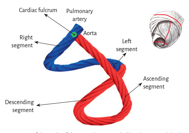
Figure 1.46. Site of the cardiac fulcrum in the myocardial band (rope model). The inset shows (in red) the schematic diagram of the rope model overlapping with the myocardium.
Conclusion. This anatomical description, both in bovid and human hearts, would put an end to the lack of support of the myocardial band to fulfill its twisting function, as the cardiac fulcrum (Figure 1.47) is the insertion point to achieve the necessary leverage, same as a muscle with its bone insertion. The observation of the tendinous cardiac fulcrum as a meeting point between the origin and end of the myocardial band would confirm its helical configuration. Moreover, by achieving energy dissipation, this nucleus would avoid the twisting motion of the ventricular helix from shifting to the aorta and hampering systolic ejection.
Конец ознакомительного фрагмента.
Текст предоставлен ООО «ЛитРес».
Прочитайте эту книгу целиком, купив полную легальную версию на ЛитРес.
Безопасно оплатить книгу можно банковской картой Visa, MasterCard, Maestro, со счета мобильного телефона, с платежного терминала, в салоне МТС или Связной, через PayPal, WebMoney, Яндекс.Деньги, QIWI Кошелек, бонусными картами или другим удобным Вам способом.