

 полная версия
A Manual of the Operations of Surgery
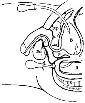
Fig. xxxvi. 160
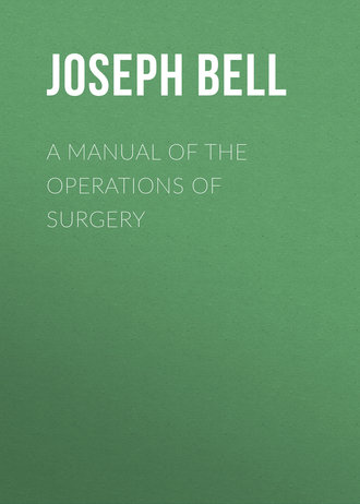
2. From the Rectum.—Except in cases of enlargement of the prostate, it is at once easier and safer to puncture the bladder from the rectum. The well-known triangular space uncovered by peritoneum, with its apex in front close to the prostate, and bounded on either side by the vasa deferentia and vesiculæ seminales, can be easily reached by a curved trocar. This should be guided by one, or, still better, by two fingers, into the rectum, with its concavity upwards, and the point should be pushed upwards by depression of the handle, whenever it is fairly behind the prostate. The trocar may then be withdrawn, and the canula retained for at least forty-eight hours by a suitable bandage. Mr. Cock, of Guy's Hospital, had a special canula for the purpose, which expands at its extremity after its introduction, and thus is not apt to slip.161 Some surgeons insist that the surgeon should be able to ascertain the existence of fluctuation between the finger in the rectum, and the other hand above the pubes. This is exceedingly difficult to elicit when the bladder is very much distended, and from the constrained position of the finger in the bowel.
Phymosis.—Elongation of the prepuce, with contraction of its orifice, in most cases congenital, sometimes so extreme as to cause difficulty in micturition, and frequently preventing the uncovering of the glans.
Operation.—In all well-marked cases, the following is required:—The elongated prepuce should be pulled forwards by a pair of catch-forceps, and a circle of skin and mucous membrane removed by a single stroke of a bistoury, or by sharp scissors. Care should be taken lest the glans be included in the incision, as has happened in at least one instance. The skin will then be found to retract very freely beyond the glans, but the mucous membrane is found still to cover the glans, and its orifice is still constricted. It must then be slit up (Fig. xxxvii. b b) on the dorsum of the glans, with probe-pointed scissors, as far as the corona, and the glans will then be thoroughly exposed. The edges of mucous membrane and skin should then be stitched to each other by at least five or six fine silk sutures, any bleeding points having been first carefully secured. The angles will in time round off, and a wonderfully seemly prepuce be obtained. This operation may be done as a method of cure for obstinate enuresis in cases in which the prepuce is very long and redundant, even when it is not too tight. The author has done this in more than twenty cases with excellent results.
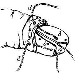
Fig. xxxvii. 162
Varieties.—When the prepuce is narrowed at its orifice without being redundant in length, a milder operation will prove sufficient. The principle is the same as in the former, but the amount of incision is less, and nothing is removed. Two methods are possible:—
1. By scissors.—The blunt point of a pair of scissors is introduced through the preputial orifice, the other blade being outside, and the skin and mucous membrane are divided for about half an inch; the skin being then retracted, the mucous membrane is still further divided by one or two additional snips, and then the edges of skin and mucous membrane are stitched together by one or two points of suture.
2. By knife.—A director being introduced within the prepuce, a narrow-bladed knife is guided along it, and pushed through the prepuce from within, and then made to divide skin and mucous membrane from within outwards. Stitches as before.
N.B.—Be careful lest the director pass into the meatus urinarius, and the glans be split up.
Again, some surgeons prefer two lateral incisions instead of one dorsal one. In this case skin and mucous membrane should be divided by scissors for about a quarter of an inch, and then a single stitch inserted in the angle of junction. This has been further modified by Cullerier, who proposed the division of the tight mucous membrane only, in three or four points. He used a pair of scissors with one sharp and one probe-pointed blade, the sharp one thrust in between skin and mucous membrane, the blunt one between the mucous membrane and the glans.
Amputation of the Penis.—This exceedingly simple operation is performed by a single stroke of an amputating knife, drawn along from heel to point, while the penis is stretched in the operator's left hand. As there is more risk of redundancy than of deficiency of the skin, no attempt is made to save it. Numerous vessels in the corpora cavernosa require ligature. Amputation of the penis may be done bloodlessly by the thermo-cautery even close to its root. Transfix the root of corpora cavernosa by a needle; above this pass two or three turns of an elastic ligature; then slowly divide at a low red heat the skin and corpora cavernosa below the needles; split the urethra after dividing its mucous membrane with a knife. The author has done this several times with ease and rapid healing.

Fig. xxxviii. 163
The chief risk is stricture of the orifice of the urethra. To prevent this, several modifications of the operation have been introduced.
1. Ricord's method.164—After the amputation the surgeon seizes with forceps the mucous membrane of the urethra, and with a pair of scissors makes four slits in it, so as to form four equal flaps, and with a silk ligature stitches each of these to the skin. Contraction of the cicatrix will thus tend to open rather than close the urethral orifice.
2. Teale's method.165—He slits up, by a bistoury on a director, the urethra and skin over it for about two-thirds of an inch, and then stitches the one to the other, thus making it a long oval dependent orifice (Fig. xxxviii.).
3. Miller's proposed method.166—"A narrow-bladed knife is first used to transfix the penis between the spongy and cavernous bodies close to the root; the knife having been carried forwards for an inch and a half, its edge is turned perpendicularly downwards, and the urethra and skin flap are divided, the cavernous bodies and dorsal integument being then cut perpendicularly upwards where the knife was originally entered for transfixion. A button-hole is afterwards made in the lower flap, though which the corpus spongiosum and urethra protrude, while the flap itself is turned upwards, and attached dorsally and laterally, so as to cover in the exposed cavernous structure."
Hydrocele.—The very simple operation necessary for hydrocele is thus performed:—The surgeon supports the tumour in his left hand so as to project it forwards, and make the scrotum as tense as possible in front. Having carefully ascertained the exact position of the testicle, which can generally be easily enough done by a finger accustomed to discriminate the difference between a soft solid, and a bag tensely filled with fluid, aided by the peculiar sensation of the testicle when squeezed, the surgeon enters a trocar and canula about an eighth of an inch in diameter into the distended cavity of the tunica vaginalis, near the fundus of the swelling. When it is evident the instrument is fairly entered, and not till then, the trocar is withdrawn, and the fluid allowed completely to drain off. When it ceases to flow the surgeon places his forefinger over the end of the canula to prevent the entrance of air, till he fits into its orifice a suitable syringe containing two drachms of the tincture of iodine, made according to the Edinburgh Pharmacopœia: the tincture of the British Pharmacopœia is not sufficiently strong. Having injected this cautiously into the cavity, the canula is withdrawn, and the surgeon, seizing the now flaccid scrotum in his right hand, gives it a thorough shake, so as to spread the iodine over as much as possible of the inner wall. When properly performed this very simple procedure very rarely fails to produce a radical cure; though less thorough operations, such as mere evacuation of the fluid, less stimulating injections, unguents introduced on probes, and the like, often fail of success, and thus give encouragement to absurdities, such as wire-setons, or to more severe operations, such as laying open the sac.
Hæmatocele.—When the contents of the sac of the tunica vaginalis are found to be grumous instead of simply serous, or when, as often happens, only pure blood escapes when the fluid is nearly evacuated, it is found that simple evacuation and injection are very rarely sufficient to effect a cure.
After they have been fairly tried, the sac of the hæmatocele should be laid open in its full extent; any large vessels which bleed should be tied, and the cavity then stuffed with lint. When the lint can be removed, which will be after two or three days, the edges of the wound should be brought closely together, and the cavity will then rapidly heal up from the bottom, and be obliterated by secondary union of granulations.
In cases where the walls of the cavity are enormously thickened, or even, as sometimes happens, almost bony in consistence, an elliptical portion may be removed with advantage.
Excision of Testicle.—This operation is rarely required except for tumours of the testicle. Hence the size of the incision necessary must vary much with the size of the tumour; and the amount of skin to be removed (if any) on the amount of adhesions it has formed to the tumour.
One or two points must be attended to in every case of extirpation of a testicle:—
1. The incision should commence over the cord just outside of the external ring, and be continued fairly over the tumour to its base.
2. As to removal of skin, some surgeons advise that none should be taken away, others that a considerable quantity can be spared. There is certainly less risk of secondary hæmorrhage if a portion be removed, than when a flaccid empty bag is left. The author invariably removes a very large quantity of skin if the tumour is large, as there is much more rapid healing, and the resulting scrotum is much more comfortable for the patient.
3. The cord should be exposed at the beginning of the operation, raised from its bed and given to an assistant, who should compress it gently, not from any fear of its escape into the abdomen, but to prevent hæmorrhage. If the tumour has been very large and heavy, the cord will have been much stretched, and if divided too high up, may really give trouble by its elasticity, unless the above precaution is taken. The cord then having been divided close to the tumour, the latter is removed, care being taken not to include the sound testicle in the removal. All the vessels are then to be tied or twisted, and the spermatic artery is to be secured alone, not, as used to be the case, included in a common ligature with the other constituents of the cord. Secondary hæmorrhage is very apt to occur from small scrotal branches which may have escaped notice during the operation.
Operations on the Anus and its Neighbourhood.—Fistula in Ano.—While much might be written on the pathology of fistula, and a good deal even on its diagnosis, a very few words will suffice to describe the simple and effectual operation for its relief.
Dismissing at once all so-called palliatives, drugs, unguents, pressure, and injections, as mere waste of time, and holding that the only method of cure consists in laying the fistula fairly open, the question narrows itself into this: What is the best method of laying it open? Prior to the discovery by Ribes of the great principle that the internal orifice of the sinus is always within an inch or an inch and a half of the orifice of the anus, the operations for fistula were most unnecessarily severe; the gut used to be divided as far up as the sinuses extended; and large portions of the anus used to be excised bodily along with the sinuses. It is now a much simpler and more satisfactory operation.
Operation.—A common silver probe bent to the required shape is passed into the external opening, or, if there are more than one, into the largest and oldest one. The forefinger of the left hand being introduced into the rectum, the probe is passed through the internal orifice, and its point brought out by the anus. The portion of tissue raised by the probe can then be easily divided with the certainty that the fistula is laid fully open. Anal fistulæ have been divided by the elastic ligature, but it seems slower in action and more painful, with no counterbalancing advantages.
The author has for last few years operated almost exclusively by a long knife which is continued into a steel probe. The probe is passed up the fistula, then into the bowel, and is hooked out at the anus, and in being simply pushed on the knife cuts the fistula—tuto, cito, et jucunde, the patient rarely knowing that more has been done than an exploration.
In cases where, from the hardness and density of the parts it is impossible to pass the probe and bring it out at the anus, a strong probe-pointed bistoury may be passed in by the external orifice till its probe-point can be felt by the finger in the bowel at the internal opening. Supported by the finger it can then be made to cut outwards till the whole septum is divided.
Fissure of the Anus, Ulcer of the Anus, resemble each other alike in the exceeding annoyance which they give to the sufferer, and in the simplicity of the treatment needed.
Operation.—Once the presence of either is determined by the finger in the anus, a sharp-pointed curved bistoury should be introduced, transfixing the base of the fissure or ulcer, and then guided on the finger, completely dividing it, so as to change the ragged ulceration into a simple wound which will rapidly heal.
Prolapsus Ani, Operation for.—Complete prolapsus in which the whole gut is involved, as seen in the very young and the very aged, is suited for palliative rather than radical treatment.
Cases of prolapsus of the mucous membrane only, as is not uncommon in connection with or as a result of hæmorrhoids in adults, give opportunity for operative interference.
We may act on either the skin or mucous membrane, or both at once.
1. The skin is often found loose, and arranged in radiating folds round the anus. In such cases, as recommended first by Dupuytren, some of these projecting folds may be removed. Again it may be prolapsed in a great loose ring or circular fold round the margin, forming an exaggerated external pile; in such a case the loose fold may be fairly excised with curved scissors, as recommended by Hey of Leeds.
The first of these methods is apt to be insufficient, the second again has the risk of removing too much.
2. If the protrusion is chiefly mucous membrane exposed in folds, or a ring, which is generally outside, one of two methods of treatment may be tried:—
a. By ligature, as recommended by Mr. Copeland. Raising a longitudinal fold of the mucous membrane, he passed a ligature round it as if it were a pile. There is less chance of the ligature slipping if a double thread be used and its base thus transfixed. Three, four, or even more folds may be thus treated.
b. When the mucous membrane has been so long exposed as to have lost many of its characters, and to resemble leather in its toughness, excision will be found less painful and much more rapid than ligature.
A longitudinal fold at each side of the anus should be pinched up and excised by a pair of probe-pointed curved scissors. There is always a certain amount of risk of hæmorrhage following such an operation. The risk is lessened and the result improved by stitching up the wound in the mucous membrane before the protruded portion of bowel is returned.
Polypi of the Rectum.—Pedunculated growths varying in consistence, shape, and size, but resembling each other in having a distinct stalk, and in frequently being protruded at stool.
Operation.—Invariably by ligature, which may be single round the stalk, if the tumour be globular and with a distinct narrow stalk, or by transfixion, if (as sometimes happens) the tumour be of uniform thickness throughout, like a worm.
Hæmorrhoids Or Piles.—In the treatment of piles it is the differential diagnosis that is troublesome and occasionally difficult; the operative interference required is generally very simple, if the nature of the case be rightly determined.
External piles.—Operation.—The apex of the soft flabby excrescence should be seized by a pair of catch-forceps, and it should be cut off close to its base with a knife, or, what is better, a pair of curved scissors. Any little vessel which jets may then be secured. If, instead of numerous individual tumours, a ring of skin round the anus be involved, the whole of it should be shaved off, but not very close to its base, lest too great contraction of the anal orifice should ensue.
If the surgeon, after excising a pile or piles, will take the trouble to stitch up the wound with catgut, he will find the cure much more rapid and less painful than when this is omitted.
Internal piles.—Incision is extremely dangerous, from the vascularity of the parts, and their being so inaccessible from their position within the sphincter ani. Hence ligature is safer and equally effectual. The patient should be directed to sit over hot water, and strain till the whole of his piles are fairly protruded. The surgeon should then transfix the base of each separately with a curved needle bearing a strong double thread. The needle being cut off, the threads should be very firmly tied, each isolating its own half of the pile. The tying should be exceedingly tight, so as to cause instant and complete strangulation and death of the tumours. All the piles should be tied at the same sitting. If the piles are very small they may be secured without transfixion in a single noose after being seized by a hook or forceps. There is greater risk of the noose slipping than when the base has been transfixed.
The strangulated masses must then be returned into the bowel, and the patient kept in bed or on a sofa till the ligatures separate, which is generally not till the fourth or fifth day. A certain amount of urinary irritation, showing itself sometimes in strangury, sometimes in complete retention, occasionally follows this operation.
Mr. Smith of King's College, and many other surgeons, treat internal piles by means of an ivory clamp to hold them tight, while they are burned off by the actual cautery or the thermo-cautery at a low red heat. They claim that pyæmia more rarely follows this mode.
There are certain cases in which the lower inch or two of the rectum are found red and congested, and in which every stool is followed by the loss of a certain quantity of florid arterial blood, and yet no distinct hæmorrhoidal tumour is to be seen. In such cases the ligature is not applicable, and relief is obtained by the application of pure nitric acid, or other potential caustics to the bleeding surface, as recommended by Houston, Lee, Smith, Ashton, and others. These cases are comparatively rare, and whenever they can be applied, the ligature is much simpler, safer, and more certain.
Venous piles.—When a sudden effusion of blood has occurred into one of the varicose veins or sinuses of a congested anus, an oval or rounded tumour is felt, very tense, shining, and painful. To slit it freely up with an abscess lancet, and evert the clot inside, at once relieves all the symptoms.
CHAPTER XIII.
TENOTOMY
For convenience' sake I group under this one head certain operations used for the relief of distortion, in which muscles or tendons are divided subcutaneously. Since the discovery of the principle by Delpech, and the application of it by Stromeyer, Dieffenbach, Little, and countless successors, it has been used for very many cases for which it is totally inapplicable, e.g. for the division of the muscles of the back in spinal curvature. Still there remain several deformities for the relief of which subcutaneous tenotomy is a most important remedy; chief among these are Wry Neck and Club-foot.
Operation for Wry Neck.—Subcutaneous section of the sterno-mastoid.—In what cases of wry neck is this operation suitable? In those only in which the muscles are the starting-point of the mischief. These are sometimes congenital, more frequently they commence in childhood. In cases where the distortion depends on disease of the cervical vertebræ, or is secondary to curvature of the spine, division of the muscle is worse than useless.
Operation.—A tenotomy knife, which should be sharp-pointed, narrow in the blade, with a blunt back, should be introduced through the skin a little to one side of the sternal portion of the affected muscle, passed along with its flat edge between the skin and the tendon, till it has fairly crossed the tendon; the blade should then be turned so that by a gradual sawing motion the edge may be made to divide the tendon about an inch above the sternum. A distinct snap will then be felt or heard, and the position of the head will be at once much improved. Exercise, warm bathing, and rubbing, will generally suffice to complete the cure, without it being necessary to call in the aid of the instrument-maker with his expensive apparatus.167
Operations for Club-Foot.—The following are the tendons which may require division in the cure of club-foot, and the operations for their division.
1. The tendo Achillis.—There are very few cases of true club-foot which can be successfully treated without division of the tendo Achillis. While in talipes equinis it is generally the only disturbing agent, in talipes varus and valgus it invariably increases and maintains the deformity, which the tibiales or peronei seem to originate.
Operation.—The foot being held at about a right angle with the leg, the operator should pinch up the skin over the tendon, introduce the knife flatwise, a little to one side of the tendon, till its point is nearly projecting at the other, then turn the edge on the tendon and cut inwards with a sawing motion till the tendon gives way with a distinct snap, and the foot can be completely flexed with ease.
Dr. Little168 recommends that the tendon should be divided from before backwards. There is more risk by this method of wounding the skin, and thus losing the subcutaneous character of the operation.
Professor Pancoast169 divides the inferior portion of the soleus muscle instead of the tendo Achillis.
2. Tibialis posticus.—Next in frequency and importance to that of the tendo Achillis, division of this tendon is much more difficult to perform. It may be performed either above or below the ankle.
(a.) Above the ankle.—The blade of a tenotomy knife should be entered perpendicularly at the posterior margin of the tibia, half an inch or an inch above the internal malleolus, so as to pass between the bone and the tendon of the tibialis posticus, the blade directed towards the latter; the assistant should now evert the foot, the operator pressing the blade against the tendon.170
(b.) Below the ankle, close to the attachment to the scaphoid. This is the better position of the two when the position of the tendon can be made out, which is not always the case, especially in cases of old standing.
Raising the skin just over the astragalo-scaphoid joint, the knife should be entered with its blade downwards, and across the tendon, and should be made to cut on the bone, while an assistant everts the foot till the tendon gives way with a distinct snap.
3. Tibialis anticus may in like manner be divided either just above the ankle, or at its insertion. When it requires division it can generally be made so prominent as to render its division comparatively easy.
4. Peronei.—These do not often require division, cases of talipes valgus being usually paralytic in character. If necessary they can be cut as they cross the fibula.
5. The plantar fascia, may require division; when this is the case, it is so prominent as to render the operation very easy, if conducted on the principles mentioned above.