

 полная версия
A Manual of the Operations of Surgery
It is rarely necessary to remove more of the bone than merely its articular extremity (when performed for disease of the joint), and if possible this should be done inside the capsule, i.e. through an incision in the capsule, but without involving its attachment to the neck of the bone. When the glenoid is also diseased, mere gouging or scraping the cartilaginous surface will not suffice, but the neck must be thoroughly exposed, so that the whole cup of the glenoid may be removed by powerful forceps.
Cases suitable for Excision.—Cases of chronic disease of the head of the humerus (generally tubercular), or of chronic ulceration of the cartilages which has resisted counter-irritation. Cases of gunshot injury of the joint, or of compound dislocation, or fracture involving the joint. Cases of limited tumours affecting merely the head and upper third of the bone, and non-malignant in character. Anchylosis very rarely requires and would not be much benefited by such an operation.
Operation.—Though perhaps not the easiest, the following method is the one followed by the best results. It is suited especially for cases of caries or other disease of the joint, where the head of the humerus is either alone or chiefly affected:—
A single straight incision (Plate I. fig. a.) is made from a point just external to the coracoid process downwards along the humerus for at least three inches. It corresponds almost exactly to the bicipital groove, and has the advantage of avoiding the great vessels and nerves. The long head of the biceps may then be raised from its groove, and drawn to a side so as to be preserved. This is deemed of importance by Langenbeck and others. Mr. Syme, however, did not attach much value to its preservation, as it is often diseased. The capsule, which is often much altered, perhaps in part destroyed, is then opened, and the tendons of the muscles which rotate the head of the humerus divided in succession, while the elbow is rotated first inwards and then outwards by an assistant so as to put them on the stretch. The arm being then forced backwards, the head of the bone can be protruded through the wound, and sawn off at the necessary distance down the shaft. The glenoid must then be carefully examined, and any diseased bone removed by the cutting pliers. One or two small branches supplying the anterior fold of the axilla are the only vessels divided, and may not even require ligature, unless, indeed, from necrosis, or to remove a tumour, a larger portion of the humerus than usual has been removed. If the limit of capsule has been infringed on below, the circumflex vessels may probably be cut, in which case the bleeding may be considerable.
N.B.—In cases of fracture of neck of humerus, or of compound gunshot injury, or where the head has been separated by necrosis from the shaft, or where, as has happened to Stanley and others, the bone broke in the endeavour to tilt the head out, the surgeon will require to seize the detached head with strong forceps, and dissect it out with care.
Other methods of Resection.—When from great thickening and induration of the soft parts, enlargement of the head of the bone, or other reason, the straight incision may be deemed insufficient for the purpose (and we may remark that there are comparatively few cases in which it is insufficient), access may be obtained to the joint by raising a flap from the deltoid (Plate III. fig. a). Its shape—V-shaped, semilunar, or ovoid—is not of much consequence, for there are no great nerves or vessels to wound on the outside of the joint, and the surgeon should be guided, as in all other operations on the joint, very much by the position of any pre-existing sinuses. This flap being raised upwards towards its base, very free access is gained to the joint.
In these cases, fortunately comparatively rare, in which there is reason to believe that the glenoid is chiefly involved in disease, and yet that the disease can be removed without amputation, access will be gained most easily by an incision (Plate III. fig. b.) on the posterior surface of the joint, corresponding in size and direction to the linear incision in front. This gives a much easier mode of access to the glenoid. I have seen this practised in one very remarkable case by Mr. Syme, in which the glenoid cavity and neck of the scapula were extensively diseased, while the head of the bone was quite sound.
After-treatment is exceedingly simple; for the first day or two the shoulder is to be supported on a pillow with a simple pad in the axilla, if there is any tendency for the arm to drag inwards; after this the patient should be encouraged to sit up and move about with his arm in a sling, the elbow hanging freely down.
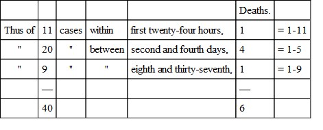
Results.—Hodge records ninety-six cases in which this excision was performed for gunshot injury, of which twenty-five proved fatal, and fifty for disease, of which only eight died,—results which are more encouraging than those of amputation at the shoulder-joint for disease; though for injury the mortality is much greater than Larrey's famous Statistics of Amputation, q.v. p. 65.
Spence had thirty-three cases, with three deaths. He generally made a counter-opening behind to get rid of discharges, and inserted a drainage-tube.
Gurlt's statistics of excision for gunshot injury give of 1661 cases 1067 recoveries, 27 doubtful results, and 567 deaths, the mortality being 34.70 per cent.
Excision of the Elbow-Joint—In what cases should it be performed?—1. For disease of the elbow-joint which has resisted ordinary remedies, and is wearing down the patient's strength, including caries, ulceration of cartilages, and gelatinous synovial degeneration.
2. For wounds of the elbow penetrating the joint, the prognosis both as to the patient's life and the usefulness of his arm is much better after excision than after endeavours to save the joint without excision. This is especially the case when the wound of the joint is small and punctured, but if the case is seen early and treated by free drainage, with antiseptic precautions, excision may not be required.
3. For anchylosis, in cases where after disease or injury the limb has stiffened in a bad position, especially when, with a straight elbow, the hand is rendered almost perfectly useless.
How much should be removed?—In the elbow-joint, more than any other joint in the body, complete excision is absolutely necessary; any portion of the articular surface being left proves a source of unfavourable result.
The surgeon is apt to err rather in removing too little than too much. For the removal of too little bone is, on the one hand, apt to result in long-standing sinuses, on the other, to induce anchylosis.
In making the section of the bones, the saw ought to be applied to the humerus transversely just at the commencement of its condyloid projections, and to the radius and ulna, at least at a level with the base of the coronoid process of the ulna.
But while removing enough, we must not be led into the error of removing too much. If this is done, as was done by Sir Philip Crampton in his first case, and as happens occasionally of necessity in cases of excision for gunshot wounds or other accidents, much of the power of the arm is lost as a consequence of the shortening and excessive mobility.
A mistaken pathology sometimes deceives in the examination of the state of the bones, and causes an unnecessary amount to be removed. For in many cases of disease the bones in the neighbourhood of the joint are stimulated to an excessive amount of what is in reality Nature's effort at repair, and while the cartilaginous surfaces are denuded of cartilage, soft, and porous, the bones close by are roughened with a stalactitic-looking growth, projecting in knobs and angles. Now, if this be mistaken for disease and removed, too much will almost certainly be taken away, and the result will be unsatisfactory.
Much less care need be taken exactly to discriminate and remove the diseased soft parts; indeed they may be left alone; the synovial membrane in a state of gelatinous degeneration sometimes presents a very formidable appearance of disease, but if the bones be properly removed, all this swelling will soon go down, and a healthy condition of parts succeed, without any clipping or paring on the surgeon's part.
Operation.—The back of the joint is of course chosen for the seat of the incisions, both because the bones are there just under the skin, and because the great vessels and nerves lie in front of the joint. The form and number of the incisions vary considerably, and ought to vary according to the nature of the case and the amount of disease or injury.
Though it is now little used, for historical interest I retain the description of the H-shaped incision (Plate III. fig. c.), used first by Moreau, and re-introduced by Mr. Syme, and used by him for most of his very numerous cases.
The posterior surface of the joint being exposed, the surgeon, with a strong straight bistoury, makes a transverse incision into the joint just above the olecranon. It should begin just far enough outside of the internal condyle to avoid the ulnar nerve, which the surgeon should protect by the forefinger of his left hand, and should extend transversely across to the outer condyle. From each end of this incision the surgeon should next make at a right angle two incisions, each about one inch and a half or two inches long, right down to the bone, thus marking out two quadrilateral flaps. These should next be raised from the bones, up and down, as much of the soft parts being retained in them as possible, so as to add to their thickness. The olecranon is thus exposed, and should be removed by saw or pliers by cutting into the greater sigmoid notch; the lateral ligaments must then be cut, if they are not already destroyed by the disease, and the humerus protruded, a proper amount of which is then to be sawn off in a transverse direction. The head of the radius is then easily removed by the bone-pliers, and the ulna also protruded, the attachment of the brachialis anticus to the coronoid process divided, and the bone sawn across just at the base of that process.
Few vessels, if any, will require ligature, and the arm being bent to nearly a right angle, the transverse incision must be very carefully sewed up with silver sutures closely set and deeply placed, as much of the future success of the joint depends on the completeness of the primary union of this incision. The external incision may also be accurately adjusted, the internal one not so completely, to allow free vent for the discharge, which is aided by the ligatures, if any are required, being brought out at its lower angle. A figure-of-8 bandage should be applied over pads of dry lint, and the limb laid on a pillow. No splint is necessary; in a few days the patient will be able to rise and walk about.
Passive motion should be begun so soon as the first inflammatory symptoms have passed off.
If properly performed, in a tolerably healthy subject, the surgeon should not be satisfied with any results short of almost perfect restoration of motion in the joint. Flexion and extension to their full extent, with a very considerable amount of pronation and supination, are to be expected, with proper care, in a patient of average intelligence.
Numerous cases are now on record where almost perfect performance of all the duties of life was retained after excision of the elbow-joint.55
In most cases it is possible, and in nearly all advisable, to excise the joint by means of a less complicated incision. Thus one long vertical incision at the posterior surface, with its centre about midway between the ulna and the external condyle, with a transverse incision at right angles to it, and reaching almost to the internal condyle, has been often practised with a very good result.
By nearly universal consent this single straight incision is now used, and when it is properly dressed and drained gives admirable results.
A single vertical incision (Plate III. fig. d.) without any transverse one, as long ago recommended by Chassaignac, is, in most cases, quite sufficient to give access. It is most suitable in cases of anchylosis, where there is little deposit of new bone, or in cases of disease of the joint, accompanied with little swelling or thickening of surrounding tissues. It has the advantage of avoiding the cicatrix of a transverse incision, which doubtless may, if at all a broad one, somewhat interfere with the future flexion of the limb, but, on the other hand, unless care is taken, it does not give such free egress for the discharge, and when there is much delay in healing, the vertical incision may leave a cicatrix nearly as troublesome as the other.
The following modification, suggested and practised by the late Mr. Maunder, seems to be a step in the right direction when it is practicable. "After a longitudinal incision crossing the point of the olecranon I next let the knife sink into the triceps muscle, and divide it longitudinally into two portions, the inner one of which is the more firmly attached to the ulna, while the outer portion is continuous with the anconeus muscle, and sends some tendinous fibres to blend with the fascia of the fore-arm. It is these latter fibres that are to be scrupulously preserved.
"Two points have to be remembered: first, the ulnar nerve, often unseen, must be lifted from its bed, and carried over the internal condyle to a safe place, and then the outer portion of the triceps muscle with its tendinous prolongation, the fascia of the fore-arm and the anconeus muscle must be dissected up, as it were, in one piece, sufficiently to allow of its being temporarily carried out over the external condyle of the humerus."56
This method aids in retaining the power of active extension of the elbow-joint.
Excision for osseous anchylosis in the extended position of the joint may be sometimes rendered very difficult by the density, firmness, and extensive hypertrophy of the bones, which become fused into one solid mass. Any attempt to isolate the bones, and remove the anchylosed joint entire, by incising the bones as if for disease, will both prove very laborious, and also probably end in doing some damage to the vessels and nerves in front. But by sawing through the anchylosis about its centre, as was pointed out many years ago by Mr. Syme, the fore-arm may be flexed, and the bones as easily displayed, cleaned, and removed, as in the operation for disease. In this operation, as there is less thickening of the skin and subjacent textures, and in consequence more risk of deficiency and even sloughing of the flaps made by the H-shaped incision, a single straight incision will serve the purpose admirably.
Partial incisions of the elbow-joint are, as a rule, less successful and more dangerous to life than complete ones, except in cases of excision for anchylosis. Even in gunshot wounds, where the bones were previously healthy, and where uninjured portions might have been left with some hopes of success, this is the case.
Dr. Heron Watson has devised the following operation for cases of anchylosis the result of injury:—(1.) A linear incision over ulnar nerve at inner side of olecranon. (2.) The ulnar nerve to be carefully turned over the inner condyle. (3.) A probe-pointed bistoury to be introduced into the elbow-joint in front of the humerus, and then behind and carried upwards, so as to divide the upper capsular attachments in front and behind. (4.) A pair of bone-forceps to be next employed to cut off the entire inner condyle and trochlea of the humerus, and then introduced in the opposite diagonal direction so as to detach the external condyle and capitulum of the humerus from the shaft. (5.) The truncated and angular end of the humerus to be divided, turned out through the incision, and smoothed across at right angles to the line of the shaft by means of the saw, whereby (6.) room might be afforded, so that partly by twisting and partly by dissection the external condyle and capitulum are removed without any division of the skin on the outer side of the arm.57 Six cases have had satisfactory results.
The mortality from this operation is considerably less than that from amputation of the arm. Of a series of excisions for disease, injury, and anchylosis, 22.15 per cent. died, while out of a similar series of amputations of the arm the mortality was 33.4 per cent.58 Our mortality of excision of the elbow here is certainly much less than the above. All of the cases, between thirty and forty, in which I have done it have recovered with but one exception, and Mr. Syme lost only one during the time I was his assistant.
Professor Spence lost only 16 in 189 cases, or 8.3 per cent.
Gurlt's statistics for gunshot injury give a mortality of over 24 per cent.
Out of 82 cases where the joint was excised for injury in the Schleswig-Holstein and Crimean campaigns, only 16 died; and out of 115 cases in which the joint was excised for disease, only 15 died.
The period after the injury at which the excision is performed seems to be important.
Excision of the Wrist.—Very various methods have been proposed and executed for the purpose of excising this joint. These vary much in difficulty and complexity, in proportion to the endeavours made to save the tendons from being cut.
The principles which must guide all attempts at operative interference with this joint are—
1. To remove all the diseased bone, including the cartilage-covered portions of the radius, ulna, and of the metacarpal bones, as little of these bones being removed as possible, beyond the cartilage-covered portions.
2. To disturb the tendons as little as possible, especially to avoid isolating them from the cellular sheath.
3. To commence passive motion of the fingers very soon after the operation.
It is rarely possible to remove the carpal bones as a whole, from the diseased condition which renders the operation necessary, and the digging out of the various bones piecemeal renders the operation very tedious, especially if the proximal ends of the metacarpal bones are involved and require to be removed, hence this operation was practically impossible till after the discovery of anæsthesia.
In describing the operation elaborated and described by Professor Lister, the type of the various plans in which the tendons are saved is given, while a very few words descriptive of the incisions used by others who cut the tendons will suffice.
Lister's Operation of Excision of the Wrist-Joint.—Even an abridgment of Mr. Lister's account of his operation must necessarily be long, because the operation itself is so complicated and prolonged, and guided by such precise principles, as to render much abridgment almost impossible.
A tourniquet is put on, to prevent oozing, which would conceal the state of the bones; any adhesions of the tendons must be then broken down by free movement of all the joints.
The radial incision (Plate IV. fig. a.) is then made. It commences at the middle of the dorsal aspect of the radius, on a level with the styloid process, passes as if going towards the inner side of the metacarpo-phalangeal joint of the thumb, in a line parallel to the extensor secundi internodii, but turns off at an angle as it passes the radial border of the second metacarpal, and then longitudinally downwards for half the length of that bone. The extensor carpi radialis brevior tendon is divided in the incision. The soft parts at the radial side are to be carefully dissected up, and the tendon of the extensor carpi radialis longior divided at its insertion. The cut tendons, and the extensor secundi internodii tendon and the radial artery can thus be pushed outwards, enabling the trapezium to be separated from the carpus by cutting-pliers. The extensor tendons being relaxed by bending back the hand, the soft parts must be cleared from the carpus as far as possible towards the ulnar side.
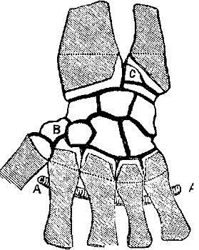
Fig. vi. 59
The ulnar incision (Plate IV. fig. b.) extends from two inches above the end of the ulna, in a line between the bone and the flexor carpi ulnaris, straight down as far as the middle of the palmar aspect of the fifth metacarpal. The dorsal lip of this incision is then raised, and the tendon of the extensor carpi ulnaris cut at its insertion, and reflected up out of its groove in the ulna along with the skin. The extensor tendons are then raised from the carpus, and the dorsal and lateral ligaments of the wrist divided, the tendons still being left as far as possible undisturbed in their relation to the radius. In front the flexor tendons are cleared from the carpus, the pisiform bone separated from the others though not removed, and the hook of the unciform divided by pliers. The knife must not go further down than the base of the metacarpal bones, in case of dividing the deep palmar arch. The anterior ligament of the wrist being now divided, the carpus and metacarpus are to be separated by cutting-pliers, and the carpus extracted by strong sequestrum forceps. By forcible eversion of the hand, the ends of radius and ulna can be protruded at the ulnar incision; as little as possible should be removed, consistent with removing all the disease. The ulna should be cut obliquely, leaving the base of the styloid process, and removing all the cartilage-covered portion. A thin slice of the radius is then to be cut also with the saw, so thin as to remove only the bevelled ungrooved portion, and leaving the tendons as far as possible undisturbed in their grooves. The ulnar articular facet is to be snipped off with bone-pliers. If the bones are more deeply carious, the diseased parts must at all hazards be removed with pliers or gouge. The metacarpal bones must then be treated in precisely the same way, their ends sawn off and their articular facets snipped off with the bone-pliers longitudinally. The trapezium is then to be seized by forceps and carefully dissected out, the metacarpal bone of the thumb pared like the others, the articular surface of the pisiform removed, the rest of the bone being left if it is sound. The radial incision is stitched closely throughout, and also the ends of the ulnar incision, any ligature being brought out through the centre of the ulnar incision, which is kept open with a piece of lint, which also gives support to the extensor tendons.
The after-treatment is important, the principal specialities being—(1.) early and free movement of the fingers; (2.) secure fixing of the wrist to procure consolidation. (1.) By passive motion of the joints of the knuckles and fingers, commenced on the second day, and continued daily after the operation; (2.) By a splint supporting the fore-arm and hand, the fingers being held in a semiflexed position by a large pad of cork fastened firmly on to the splint and made to fit the palm; this prevents the splint from slipping up the arm, and by a turn of a bandage insures fixation of the wrist-joint. The anterior part of this splint below the fingers may be gradually shortened, allowing more and more passive motion of the fingers, but the patient must wear it for months, indeed, till he finds his wrist as strong without it as with it.
Among the various operations that have been devised, the following require notice:—Mr. Spence, Dr. Gillespie, Dr. Watson, and the author, use a single dorsal incision with excellent results, and find it quite easy to remove all the bones from it. Mr. Spence had sixteen cases without a death.
Posterior Semilunar Flap, from carpal attachment of metacarpal of index finger round to styloid process of ulna; dividing integuments only, then separating the tendons of the common extensor longitudinally, and drawing them aside by blunt hooks, the diseased bones are removed piecemeal by curved parrot-bill forceps.60
Posterior Curved Flap.—An incision down to the carpal bones, extended from a point two lines to the ulnar side of the extensor secundi internodii pollicis, and from a quarter to half an inch below the radio-carpal articulation, swept in a curvilinear direction downwards, close to the carpal extremities of the metacarpal bones, to a point just below the end of the ulna. The flap thus marked out was dissected up, and consisted of the integuments, areolar tissue, and extensor tendons of the four fingers, together with large deposits of fibrine, the products of repeated and prolonged inflammatory action. The tendon of the second extensor and its soft parts around were separated from the bones. The remains of the ligaments were cut, flexion of the hand protruded the carious ends of radius and ulna. The bones were then dissected out, leaving the trapezium, which was not diseased, and hand placed on a splint.61