 полная версия
A Treatise on Domestic Economy; For the Use of Young Ladies at Home and at School
It is the universal law of the human frame, that exercise is indispensable to the health of the several parts. Thus, if a blood-vessel be tied up, so as not to be used, it shrinks, and becomes a useless string; if a muscle be condemned to inaction, it shrinks in size, and diminishes in power; and thus it is also with the bones. Inactivity produces softness, debility, and unfitness for the functions they are designed to perform. This is one of the causes of the curvature of the spine, that common and pernicious defect in the females of America. From inactivity, the bones of the spine become soft and yielding; and then, if the person is often placed, for a length of time, in positions that throw the weight of the body unequally on certain portions of the spine, they yield to this frequent compression, and a distortion ensues. The positions taken by young persons, when learning to write or draw, or to play on the guitar, harp, or piano, and the position of the body when sleeping on one side, on high pillows, all tend to produce this effect, by throwing the weight of the body unequally, and for a length of time, on particular parts of the spine.
Fig. 5.

MUSCLES
The muscles are the chief organs of motion, and consist of collections of fine fibres or strings, united in casings of membrane or thin skin. They possess an elastic power, like India rubber, which enables them to extend and contract. The red meat in animals consists of muscles. Every muscle has connected with it nerves, veins, and arteries; and those designed to move the bones, are fastened to them by tendons at their extremities. The muscles are laid over each other, and are separated by means of membranes and layers of fat, which enable them to move easily, without interfering with each other.
The figure on page 74, represents the muscles of the arm, as they appear when the skin and fat are removed. The muscles a and b are attached, at their upper ends, to the bone of the arm, and by their lower ends to the upper part of the fore arm, near the elbow joint. When the fibres of these muscles contract, the middle part of them grows larger, and the arm is bent at the elbow. The muscle c, is, in like manner, fastened, by its upper end, to the shoulder blade and the upper part of the arm, and by its lower end to one of the bones of the fore arm, near the elbow. When the arm is bent, and we wish to straighten it, it is done by contracting this muscle. The muscles d, d, are fastened at one end near the elbow joint, and at the other near the ends of the fingers; and on the back of the hand are reduced in size, appearing like strong cords. These cords are called tendons. They are employed in straightening the fingers, when the hand is shut. These tendons are confined by the ligament or band, e, which binds them down, around the wrist, and thus enables them to act more efficiently, and secures beauty of form to the limb. The muscles at f, are those which enable us to turn the hand and arm outward. Every different motion of the arm has one muscle to produce it, and another to restore the limb to its natural position. Those muscles which bend the body are called flexors; those which straighten it, extensors. When the arm is thrown up, one set of muscles is used; to pull it down, another set: when it is thrown forward, a still different set is used; when it is thrown back, another, different from the former; when the arm turns in its socket, still another set is used; and thus every different motion of the body is made by a different set of muscles. All these muscles are compactly and skilfully arranged, so as to work with perfect ease. Among them, run the arteries, veins, and nerves, which supply each muscle with blood and nervous power, as will be hereafter described. The size and strength of the muscles depend greatly on their frequent exercise. If left inactive, they grow thin and weak, instead of giving the plumpness to the figure, designed by Nature. The delicate and feeble appearance of many American women, is chiefly owing to the little use they make of their muscles. Many a pale, puny, shad-shaped girl, would have become a plump, rosy, well-formed person, if half the exercise, afforded to her brothers in the open air, had been secured to her, during childhood and youth.
NERVES
The nerves are the organs of sensation. They enable us to see, hear, feel, taste, and smell; and also combine with the bones and muscles in producing motion.
The first engraving, on p. 77, (Fig. 6,) is a vertical section of the skull, and of the spinal column, or back bone, which supports the head, and through which runs the spinal cord, whence most of the nerves originate. It is a side view, and represents the head and spine, as they would appear, if they were cut through the middle, from front to back. Fig. 7, exhibits them as they would appear, if viewed from behind. In Fig. 6, a, represents the cerebrum, or great brain; b, the cerebellum, or little brain, which is situated directly under the great brain, at the back and lower part of the head; c, d, e, is the spinal marrow, which is connected with the brain at c, and runs through the whole length of the spinal column. This column consists, as has already been stated, of a large number of small bones, f, f, called vertebræ, laid one above another, and fastened together by cartilage, or gristle, g, between them.
Fig. 6.
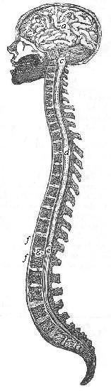
Fig. 7.
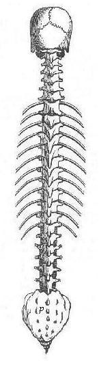
Between each two vertebræ, or spinal bones, there issues from the spine, on each side, a pair of nerves. The lower broad part of the spine, (see p, Fig. 1, p. 70, and Fig. 7, p. 77,) is called the sacrum; in this, are eight holes, through which the lower pairs of nerves pass off.
The nerves of the head and lungs run directly from the brain; those of all other parts of the body proceed from the spine, passing out in the manner already mentioned.
The nerves which thus proceed from the spine, branch out, like the limbs and twigs of a tree, till they extend over the whole body; and, so minutely are they divided and arranged, that a point, destitute of a nerve, cannot be found on the skin.
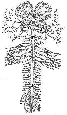
Some idea of the ramifications of the nerves, may be obtained by reference to the following engraving, (Fig. 8.) In this, A, A, represents the cerebrum, or great brain; B, B, the cerebellum, or little brain; (see also a, b, in Fig. 6;) C, C, represents the union of the fibres of the cerebrum; D, D, the union of the two sides of the cerebellum; E, E, E, the spinal marrow, which passes through the centre of the spine, (as seen at c, d, e, in Fig. 6;) 1, 2, 3, 4, 5, 6, branches of the nerves going to different parts of the body. As the nerves are the organs of sensation, all pain is an affection of some portion of the nerves. The health of the nerves depends very greatly on the exercise of the muscles, with which they are so intimately connected. This shows the reason why the headache, tic douloureux, diseases of the spine, and other nervous affections, are so common among American women. Their inactive habits, engender a debility of the nervous system, and these diseases follow, as the consequence.
Fig. 8.
It can be seen, by a reference to the side view, represented on page 77, (Fig. 6,) that the spine is naturally curved back and forward. When, from want of exercise, its bones are softened, and the muscles weakened, the spine acquires an improper curve, and the person becomes what is called crooked, having the neck projected forward, and, in some cases, having the back convex, where it should be concave. Probably one half of the American women have the head thus projecting forward, instead of carrying it in the natural, erect position, which is both graceful and dignified.
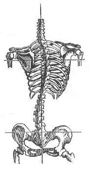
The curvature of the spine, spoken of in this work as so common, and as the cause of so many diseases among American women, is what is denominated the lateral curvature, and is much more dangerous than the other distortion. The indications of this evil, are, the projection of one shoulder blade more than the other, and, in bad cases, one shoulder being higher, and the hip on the opposite side more projecting, than the other. In this case, the spine, when viewed from behind, instead of running in a straight line, (as in Fig. 7 and 9,) is curved somewhat, as may be seen in Figures 10 and 11.
This effect is occasioned by the softness of the bones, induced by want of exercise, together with tight dressing, which tends to weaken the muscles that are thus thrown out of use. Improper and long continued positions in drawing, writing, and sleeping, which throw the weight of the body on one part of the spine, induce the same evil. This distortion is usually accompanied with some consequent disease of the nervous system, or some disarrangement of the internal organs.
By comparing Figures 9 and 11, the difference between a natural and distorted spine will be readily perceived. In Fig. 10, the curved line shows the course of the spine, occasioned by distortion; the perpendicular line, in this and Fig. 11, indicates the true direction of the spine; the horizontal lines show that one shoulder and hip are forced from their proper level.
Fig. 9.
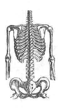
Fig. 10.
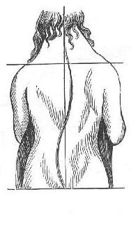
Fig. 11.
BLOOD-VESSELS
The blood is the fluid into which our food is changed, and which is employed to minister nourishment to the whole body. For this purpose, it is carried to every part of the body, by the arteries; and, after it has given out its nourishment, returns to the heart, through the veins.
The subjoined engraving, (Fig. 12,) which presents a rude outline of the vascular system, will more clearly illustrate this operation, as we shall presently show.
Fig. 12.
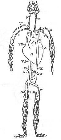
Before entering the heart, the blood receives a fresh supply of nourishment, by a duct which leads from the stomach. The arteries have their origin from the heart, in a great trunk, called the aorta, which is the parent of all the arteries, as the spinal marrow is the parent of the nerves which it sends out. When the arteries have branched out into myriads of minute vessels, the blood which is in them passes into as minute veins; and these run into each other, like the rills and branches of a river, until they are all united in two great veins, which run into the heart. One of these large receivers, called the vena cava superior, or upper vena cava, brings back the blood from the arms and head, the other, the vena cava inferior, or lower vena cava, brings back the blood from the body and lower limbs.
In the preceding figure, H, is the heart, which is divided into four compartments; two, called auricles, used for receiving the blood, and two, called ventricles, used for sending out the blood. A, is the aorta, or great artery, which sends its branches to every part of the body. In the upper part, at a, a, a, are the main branches of the aorta, which go to the head and arms. Below, at a, a, are the branches which go to the lower limbs. The branches which set off at X, X, are those by which the intestines are supplied by vessels from the aorta. Every muscle in the whole body, all the organs of the body, and the skin, are supplied by branches sent off from this great artery. When the blood is thus dispersed through any organ, in minute vessels, it is received, at their terminations, by numerous minute veins, which gradually unite, forming larger branches, till they all meet in either the upper or lower vena cava, which returns the blood to the heart. V I, is the vena cava inferior, which receives the blood from the veins of the lower parts of the body, as seen at v, v. The blood, sent into the lower limbs from the aorta, is received by minute veins, which finally unite at v, v, and thus it is emptied through the lower vena cava into the heart: o, o, represent the points of entrance of those tributaries of the vena cava, which receive that blood from the intestines, which is sent out by the aorta at X, X. In the upper part, V S, is the vena cava superior, which receives the blood from the head and arms; v, v, v, are the tributaries of the upper vena cava, which bring the blood back from the head and arms; d, d, represents the course of the thoracic duct, a delicate tube by which the chyle is carried into the blood, as mentioned on page 89; t, shows the place where this duct empties into a branch of the vena cava.
It thus appears, that wherever a branch of the aorta goes to carry blood, there will be found a tributary of the upper or lower vena cava, to bring it back.
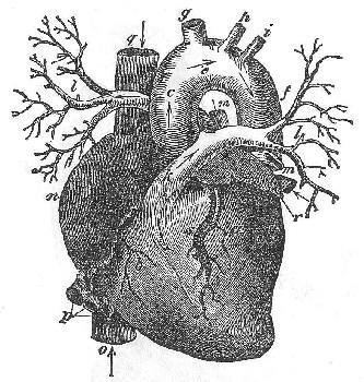
The succeeding engravings, will enable the reader to form a more definite idea of this important function of the system,—the circulation of the blood. The heart, in man, and in all warm-blooded animals, is double, having two auricles and two ventricles. In animals with cold blood, (as fishes,) the heart is single, having but one auricle and one ventricle. Fig. 13, represents the double heart as it appears when the two sides are separated, and also the great blood-vessels; those on the left of the figure being on the right side of the body, and vice versa. The direction of the blood is represented by the arrows. A, represents the lower vena cava, returning the blood from the lower parts of the body, and L, the upper vena cava, returning the blood from the head and arms. B, is the right sinus, or auricle, into which the returned blood is poured. From this cavity of the heart, the blood is carried into the right ventricle, C; and from this ventricle, the pulmonary arteries, D, convey into the lungs the blood which is returned from the body. These five vessels, A, B, C, D, and L, belong to the right side of the heart, and contain the venous or dark-colored blood, which has been through the circulation, and is now unfit for the uses of the system, till it has passed through the lungs.
Fig. 13.
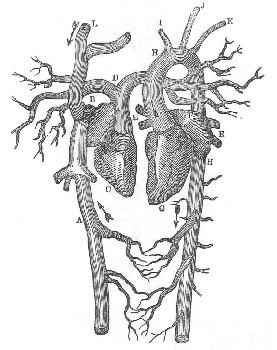
When the blood reaches the lungs, and is exposed to the action of the air which we breathe, it throws off its impurities, becomes bright in color, and is then called arterial blood. It then returns to the left side of the heart, (on the right of the engraving,) by the pulmonary veins E, E, (also seen at m, m, Fig. 15,) into the left auricle F, whence it is forced into the ventricle, G. From the left ventricle, proceeds the aorta, H, H, which is the great artery of the body, and conveys the blood to every part of the system. I, J, K, are branches of the aorta, going to the head and arms.
Fig. 14.

Fig. 14, represents the heart, with its two sides united as in nature; and will be understood from the description of Fig. 13.
On the opposite page, Fig. 15, represents the heart, with the great blood-vessels, on a still larger scale; a, being the left ventricle; b, the right ventricle; c, e, f, the aorta, or great artery, rising out of the left ventricle; g, h, i, the branches of the aorta, going to the head and arms; k, l, l, the pulmonary artery, and its branches; m, m, veins of the lungs, which bring the blood back from the lungs to the heart; n, right auricle; o, vena cava inferior; p, veins returning blood from the liver and bowels; q, the vena cava superior; r, the left auricle; s, the left coronary artery, which distributes the blood exclusively to the substance of the heart.
Fig. 15.
ORGANS OF DIGESTION AND RESPIRATION
Digestion and respiration are the processes, by which the food is converted into blood for the nourishment of the body. The engraving on the next page (Fig. 16) shows the organs by which these operations are performed.
In the lower part of the engraving, is the stomach, marked S, which receives the food through the gullet, marked G. The latter, though in the engraving it is cut off at G, in reality continues upwards to the throat. The stomach is a bag composed of muscles, nerves, and blood-vessels, united by a material similar to that which forms the skin. As soon as food enters the stomach, its nerves are excited to perform their proper function of stimulating the muscles. A muscular (called the peristaltic) motion immediately commences, by which the stomach propels its contents around the whole of its circumference, once in every three minutes.
Fig. 16.
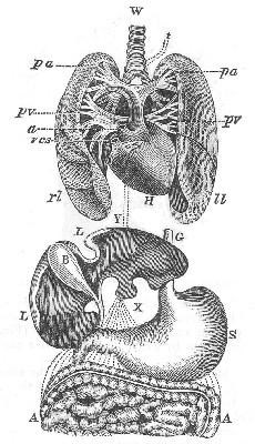
This movement of the muscles attracts the blood from other parts of the system; for the blood always hastens to administer its supplies to any organ which is called to work. The blood-vessels of the stomach are soon distended with blood, from which the gastric juice is secreted by minute vessels in the coat of the stomach. This mixes with the food, and reduces it to a soft pulpy mass, called chyme. It then passes through the lower end of the stomach, into the intestines, which are folded up in the abdomen, and the upper portion, only, of which, is shown in the engraving, at A, A. The organ marked L, L, is the liver, which, as the blood passes through its many vessels, secretes a substance called bile, which accumulates in the gall-bladder, marked B. After the food passes out of the stomach, it receives from the liver a portion of bile, and from the pancreas the pancreatic juice. The pancreas does not appear in this drawing, being concealed behind the stomach. These two liquids separate the substance which has passed from the stomach, into two different portions. One is a light liquid, very much like cream in appearance, and called chyle, of which the blood is formed; the other is a more solid substance, which contains the refuse and useless matter, with a smaller portion of nourishment; and this, after being further separated from the nourishing matter which it contains, is thrown out of the body. There are multitudes of small vessels, called lacteals, which, as these two mixed substances pass through the long and winding folds of the intestines in the abdomen, absorb the chyle, and convey it to the thoracic duct, which runs up close by the spine, and carries the chyle, thus received, into a branch of the vena cava superior, at t, whence it is mingled with the blood going into the heart. In this engraving, the lacteals and thoracic duct are not shown; but their position is indicated by the dotted lines, marked X, Y; X, being the lacteals, and Y, the thoracic duct.
In the upper half of the engraving, H represents the heart; a, the commencement of the aorta; v c s, the termination of the vena cava superior. On each side of the heart, are the lungs; l l, being the left lobe, and r l, the right lobe. They are composed of a network of air-vessels, blood-vessels, and nerves. W, represents the trachea, or windpipe, through which, the air we breathe is conducted to the lungs. It branches out into myriads of minute vessels, which are thus filled with air every time we breathe. From the heart, run the pulmonary arteries, marked p a. These enter the lungs and spread out along-side of the branches of the air-vessels, so that every air-vessel has a small artery running side by side with it. When the two vena cavas empty the blood into the heart, the latter contracts, and sends this blood, through these pulmonary arteries, into the lungs.
As the air and blood meander, side by side, through the lungs, the superabundant carbon and hydrogen of the blood combine with the oxygen of the air, forming carbonic acid gas, and water, which are thrown out of the lungs at every expiration. This is the process by which the chyle is converted into arterial blood, and the venous blood purified of its excess of carbon and hydrogen. When the blood is thus prepared, in the lungs, for its duties, it is received by the small pulmonary veins, which gradually unite, and bring the blood back to the heart, through the large pulmonary veins, marked p v, p v.
On receiving this purified blood from the lungs, the heart contracts, and sends it out again, through the aorta, to all parts of the body. It then makes another circuit through every part, ministering to the wants of all, and is afterwards again brought back by the veins to receive the fresh chyle from the stomach, and to be purified by the lungs.
The throbbing of the heart is caused by its alternate expansion and contraction, as it receives and expels the blood. With one throb, the blood is sent from the right ventricle into the lungs, and from the left ventricle into the aorta.
Every time we inspire air, the process of purifying the blood is going on; and every time we expire the air, we throw out the redundant carbon and hydrogen, taken from a portion of the blood. If the waist is compressed by tight clothing, a portion of the lungs be compressed, so that the air-vessels cannot be filled. This prevents the perfect purification and preparation of the blood, so that a part returns back to the heart unfitted for its duties. This is a slow, but sure, method, by which the constitution of many a young lady is so undermined that she becomes an early victim to disease and to the decay of beauty and strength. The want of pure air is another cause, of the debility of the female constitution. When air has been rendered impure, by the breath of several persons, or by close confinement, it does not purify the blood properly. Sleeping in close chambers, and sitting in crowded and unventilated schoolrooms, are frequent causes of debility in the constitution of young persons.
OF THE SKIN
The skin is the covering of the body, and has very important functions to perform. It is more abundantly supplied with nerves and blood-vessels than any other part; and there is no spot of the skin where the point of the finest needle would not pierce a nerve and blood-vessel. Indeed, it may be considered as composed chiefly of an interlacing of minute nerves and blood-vessels, so that it is supposed there is more nervous matter in the skin, than in all the rest of the body united, and that the greater portion of the blood flows through the skin.
The whole animal system is in a state of continual change and renovation. Food is constantly taken into the stomach, only a portion of which is fitted for the supply of the blood. All the rest has to be thrown out of the system, by various organs designed for this purpose. These organs are,—the lungs, which throw off a portion of useless matter when the blood is purified; the kidneys, which secrete liquids that pass into the bladder, and are thrown out from the body by that organ; and the intestines, which carry off the useless and more solid parts of the food, after the lacteals have drawn off the chyle. In addition to these organs, the skin has a similar duty to perform; and as it has so much larger a supply of blood, it is the chief organ in relieving the body of the useless and noxious parts of the materials which are taken for food.