полная версия
Common Objects of the Microscope
One of the tubes or channels by which these glands are enabled to pour their contents to the outside of the body, and, if they be kept perfectly clean, to disperse them into the air, is seen running up the centre of the figure, and terminating in a cup-shaped orifice on the surface of the cuticle. On the palm of the hand very nearly three thousand of these ducts lie within the compass of a square inch, and more than a thousand in every square inch of the arm and other portions of the body, so that the multitude of these valuable organs may be well estimated, together with the absolute necessity for keeping the skin perfectly clean in order to enjoy full health.
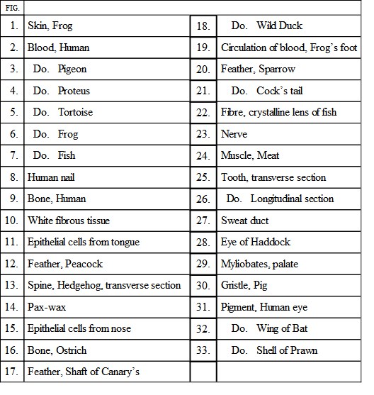
Fig. 1 shows a specimen of epidermis taken from the skin of a frog, exhibiting the flattened cells which constitute that structure, and the oval or slightly elongated nuclei, of which each cell has one. In Fig. 32, a portion of a bat’s wing, the arrangement of the pigment is remarkably pretty. Immediately above, at Fig. 31, is some of the pigment taken from the back of the human eye-ball. The shape of the pigment cells is well shown. Similar specimens may easily be obtained from the back of a sheep’s eye which has been hardened in spirit, or from that of a boiled fish. Fig. 33 shows the pigment in the shell of the prawn.
On various parts of animal structures, such as the lining of internal cavities, the interior of the mouth, and other similar portions of the body, the cells are developed into a special form, which is called “Epithélium,” and which corresponds to the epidermis of the exterior surface of the body. The cells which form this substance are of different shapes, according to their locality. On the tongue, for example (for which see Fig. 11), they are flattened, and exhibit their nucleus, in which the nucléolus may be discovered with a little care. Cells of this kind are rounded, as in the case just mentioned, or angular, and in either case they are termed squamous (i.e., scaly) epithelium. Sometimes they are like a number of cylinders, cones, or pyramids, ranged closely together, and are then called cylindrical epithelium. Sometimes the free ends of cylindrical epithelium are furnished with a number of vibrating filaments or cilia, and in this case the structure is called “ciliated” epithelium. Cylindrical epithelium may be found in the ducts of the glands which open into the intestines, as well as in the glands that secrete tears; and ciliated epithelium is seen largely in the windpipe, the interior of the nose, etc. A specimen taken from the nose is seen at Fig. 15. A beautiful example of ciliated epithelium is to be found in the gills of the mussel. A portion of one of the yellowish bands which lie along the edge of the shell on the opening side is carefully removed with sharp scissors, and examined in the shell-liquor, being protected from pressure by placing a piece of paper beneath each end of the cover-glass. Such a preparation is shown in Plate IX. Fig. 39, but no drawing can give an idea of its wonderful beauty and interest. The cilia will continue to move for a long time after removal from the shell.
Bone in its various stages is figured on Plate X.
Fig. 9 is a good example of human bone, and is a thin transverse section taken from the thigh. When cut across, bone exhibits a whitish structure filled with little dottings that become more numerous towards the centre, and are almost invisible towards the circumference. In the centre of the bone there is a cavity, which contains marrow in the mammalia and air in the birds. When placed under a microscope, bone presents the appearance shown in the illustration.
The large aperture in the centre is one of innumerable tubes that run along the bone, and serve to allow a passage to the vessels which convey blood from one part of the bone to another. They are technically called Haversian canals, and if a longitudinal section be made they will be found running tolerably parallel, and communicating freely with each other. Around each Haversian canal may be seen a number of little black spots with lines radiating in all directions, and looking something like flattened insects. These are termed bone-cells or “lacúnæ,” and the little black lines are called “canalículi.” In the living state they contain cells which are concerned in the growth of the bone, and these may be made evident by softening fresh bone with acid, cutting sections of it, and staining. When viewed by transmitted light the lacunæ and canaliculi are black; but when seen by dark-field illumination the Haversian canals become black, and the lacunæ are white.
As these canaliculi exist equally in every direction, it is impossible to make a section of bone without cutting myriads of them across; and when a high power is employed they look like little dots scattered over the surface. A very pretty object can be made of the bone taken from a young animal which has been fed with madder, as the colour gets into the bone and settles chiefly round the Haversian canal. A young pig is a very good subject, so is a rabbit.
Fig. 16 is a similar section cut from the leg-bone of an ostrich.
The development of bone is beautifully shown in Fig. 30, a delicate slice taken from a pig’s rib. Above may be seen the gristle or cartilage, with the numerous rows of cells; below is the formed bone, with one of the Haversian canals and its contents; while between the two may be seen the cartilage-cells gathering together and arranging themselves into form. The cartilage-cells are well shown in Fig. 28, which is a portion of the cup which had contained the eye of a haddock.
The horn-like substances at the end of our fingers, which we call the nails, are composed of innumerable flattened cells. These cells are generally so fused together as to be quite indistinguishable even with a microscope, but can be rendered visible by soaking a section of nail in liquor potassæ, which causes the cells to swell up and resume to a degree their original rounded form.
It is worthy of remark that the animal form is built up of cells, as is the case with the vegetables, although the cells are not so variable in shape. They generally may be found to contain well-marked nuclei, two or more of the latter being often found within a single cell, and in many cases the tiny nucleoli are also visible. Good examples of these cells may be obtained from the yolk of an egg, and by careful management they may be traced throughout every part of the animal form.
The teeth have many of the constituents of bone, and in some of their parts are made after precisely the same fashion. When cut, the teeth are seen to consist of a hard substance, called enamel, which coats their upper surfaces, of dentine, or ivory, within the enamel, and of “cement,” which surrounds the fangs. In Fig. 26, Plate X., which is a longitudinal section of the human “eye” tooth, is seen the ivory occupying the greater part of the tooth, coated by the enamel at the top and the cement at the bottom. In the centre of each tooth there is a cavity, which is plentifully filled with a pulpy substance by which the tooth is nourished, and which conveys the nerves which endow it with sensation. A traverse section of the same tooth is seen in Fig. 25.
The enamel is made of little elongated prisms, all pointing to the centre of the tooth. When viewed transversely, their ends are of a somewhat hexagonal shape, something like an irregular honeycomb. The dentine is composed of a substance pierced with myriads of minute tubes. They require a rather high power—say 300 diameters—to show them properly. The cement is found at the root of the fangs, and is best shown in the tooth of an aged individual, when it assumes very clearly the character of bone.
Sections may be made by sawing a slice in the required direction, polishing one side, and cementing it with old Canada balsam to a slide. It may then be filed down to nearly the required thinness, finished by carefully rubbing with a hone, and polished with buff leather. Canada balsam may then be dropped upon it, and a glass cover pressed firmly down.
Sections of young bone form magnificent objects for the polariser.
Fig. 29 is a section cut from one of the palate teeth of the ray (Myliobátes).
A rather important element in the structure of animals is the “elastic ligament,” which is found in the back of the neck and other parts of the body, especially about the spine. It is made of a vast number of fibres of variable shape and length, branching and communicating, arranged generally in bundles, and remarkable for containing very few vessels, and no nerves at all. At Fig. 14 may be seen an example of elastic ligament, popularly called “paxwax,” taken from the neck of a sheep.
The white fibrous tissue by which all the parts of the body are bound together is seen at Fig. 10; and at Fig. 11 is a beautiful example of the “ultimate fibres” of the crystalline lens of a sturgeon’s eye.
The muscles of animals are of two kinds, the one termed the striped, and the other the unstriped. Of these, the latter belongs to organs which work independently of will, such as the stomach, etc., while the former belongs to those portions of the body which are subject to voluntary motion, such as the arm and the leg. The unstriped muscle is very simple, consisting merely of long spindle-shaped cells, but the striped or voluntary muscle is of more complex construction. Every voluntary muscle consists of myriads of tiny fibres, bound together in little bundles, enveloped in a kind of sheath. Fig. 24 is an example of this muscular fibre, taken from beef. When soaked in spirit, it often splits into a number of discs, the edges of which are marked by the transverse lines.
A fibre of nerve is drawn at Fig. 23, and is given for the purpose of showing the manner in which the nerve is contained in and protected by its sheath, just like a telegraph-wire in its coverings. Just above is a transverse section of the same fibre, showing the same arrangement from another point of view, and also illustrating the curious phenomenon, that when nerve-fibres are treated with carmine the centre takes up the colouring matter, while the sheath remains white as before. The best way of studying nerves is to decapitate a frog, and cut off a piece of one of the nerves, which, like fine silk threads, come out between the joints of the spine inside the abdomen. By careful teasing out it is easy to obtain preparations showing all the above points, and, in addition, the folding-in of the internal sheath which correspond to the insulators of a telegraph-line.
The blood of animals is analogous in its office to the sap of plants, but differs greatly from it under the microscope. In sap there seem to be no microscopic characters, except that when a branch is cut, as in the vine, the flowing sap may contain certain substances formed in the wounded cells, such as chlorophyll, starch, and raphides; but the blood is known to be an exceedingly complex substance both in a microscopic and a chemical point of view. When a little fresh blood is placed under the microscope, it is seen to consist of a colourless fluid filled with numerous little bodies, commonly called “blood-globules,” varying very greatly in size and shape, according to the animal from which they were taken. Those of the reptiles are very large, as may be seen at Fig. 4, Plate X., which represents a blood corpuscle of the Proteus. In this curious reptile the globules are so large that they may be distinguished during its life by means of a common pocket lens.
In the vertebrated animals these corpuscles are red, and give to the blood its peculiar tint. They are accompanied by certain colourless corpuscles, spherical in form, which are sometimes, as in man, larger than the red globules, and in others, as in the siren and the newt, considerably smaller. The general view of the red corpuscles has sufficient character to enable the practised observer to name the class of animal from which it was taken, and in some cases they are so distinctive that even the genus can be ascertained with tolerable certainty. In point of size, the reptiles have the largest and the mammalia the smallest, those of the Proteus and the musk-deer being perhaps the most decidedly opposed to each other in this respect.
IX.

IX.
In shape, those of the mammalia are circular discs, mostly with a concave centre, though the camel has oval ones; those of the birds are more or less oval and convex; those of the reptiles are decidedly oval, very thin, and have the nucleus projecting; and those of the fishes are oval and mostly convex. During the process of coagulation the blood corpuscles run together into a series of rows, just as if a heap of pence had been piled on each other and then pushed down, so that each penny overlaps its next neighbour.
These objects are illustrated by six examples on Plate X. Fig. 2 is human blood, showing one of the white corpuscles; Fig. 3 is the blood of the pigeon; Fig. 4, of the Proteus anguínus; Fig. 5, of the tortoise; Fig. 6, of the frog, showing the projecting nucleus; and Fig. 7, of the roach. The blood possesses many curious properties, which cannot be described in these few and simple pages.
In the centre of Plate X. is a large circular figure representing the membrane of a frog’s foot as seen through the microscope, and exhibiting the circulation of the blood. The mode of arranging the foot so as to exhibit the object without hurting the frog is simple enough.
Take an oblong slip of wood,—my own was made in five minutes out of the top of a cigar-box,—bore a hole about an inch in diameter near one end, and cut a number of little slits all round the edge of the wooden slip. Then get a small linen bag, put the frog into it, and dip him into water to keep him comfortable. When he is wanted, pull one of his hind feet out of the bag, draw the neck tight enough to prevent him from pulling his foot back again, but not sufficiently tight to stop the circulation. Have a tape fastened to the end of the bag, and tie it down to the wooden slide. Then fasten a thread to each of his toes, bring the foot well over the centre of the hole, stretch the toes well apart, and keep them in their places by hitching the threads into the notches on the edge of the wooden strip. Perhaps an easier plan is to secure the threads by drops of sealing-wax when in the desired position. Push a glass slide carefully between the foot and the wood, so as to let the membrane rest upon the glass, and be careful to keep it well wetted. If the frog kick, as he will most likely do, pass a thin tape over the middle of the leg, and tie him gently down to the slide.
Bring the glass into focus, and the foot will present the appearance so well depicted in the engraving. The veins and arteries are seen spreading over the whole of the membrane, the larger arteries being often accompanied by a nerve, as seen in the illustration. Through all these channels the blood continually pours with a rather irregular motion, caused most probably by the peculiar position of the reptile. It is a most wonderful sight, of which the observer is never tired, and which seems almost more interesting every time that it is beheld.
The corpuscles go pushing and jostling one another in the oddest fashion, just like a British crowd entering an exhibition, each one seeming to be elbowing its way to the best place. To see them turning the corners is very amusing, for they always seem as if they never could get round the smaller vessels, and yet invariably accomplish the task with perfect ease, turning about and steering themselves as if possessed of volition, and insinuating their ends when they could not pass crosswise.
By putting various substances, such as spirit or salt, upon the foot, the rapidity of the circulation at the spot can be greatly increased or reduced at will, or even stopped altogether for a while, and the phenomenon of inflammation and its gradual natural cure be beautifully illustrated. The numerous black spots upon the surface are pigment-cells.
The tails of young fish also afford excellent objects under the microscope, as the circulation can be seen nearly as well as in the frog’s foot. The gills of tadpoles can also be arranged upon the stage with a little care, and the same organs in the young of the common newt will also exhibit the circulation in a favourable manner. The frog, however, is perhaps the best, as it can be arranged on the “frog-plate” without difficulty, and the creature may be kept for months by placing it in a cool, damp spot, and feeding it with flies, little slugs, and similar creatures.
CHAPTER VIII
Pond-Life—Apparatus and Instructions for Collecting Objects—Methods of Examination—Sponge—Infusoria.
Of all departments of microscopic research the most fascinating and the most popular is that which deals with what is known by the generic name of “pond-life.” The minute forms of the animal creation included in this term are of such exquisite beauty, and allow the processes of their life-history to be followed with such facility, from the cradle (when they have one) to the grave (which is very often the body of another, larger, organism), that there is none which has attracted more observers. Indeed, the first application of the microscope, by Leeuwenhoek, early in the seventeenth century, was to the observation of these forms of life.
X.

X.
A few words may be said, in the first place, as to the outfit. A very useful part of it is a walking-stick, to which can be attached either a net for capturing the larger forms of life, or a hook for collecting the weeds, to which many forms of great interest and beauty are attached (Fig. 15). The stick is telescopic, and can also have attached to it a bottle, which, put into the water at any desired spot,—say, amongst a clump of weeds, or near the bottom, upside down, and then suddenly reversed,—will bring away samples of the inhabitants of the neighbourhood. When these are sparsely distributed through the water, the latter may be concentrated by the use of a bottle round the neck of which is firmly tied a coarse calico bag, funnel-shaped, and supported by a wire ring, somewhat as shown in the illustration. Muslin is, however, too coarse for many organisms. This net is immersed in the water so that the ring is just above the surface, and one bottleful after another poured through. The water strains off, the organisms are left behind. The immersion is necessary to reduce the pressure to which delicate organisms would otherwise be subjected. When the bottle is full, or sufficiently concentrated as to its contents, the latter are poured into one of the ordinary collecting-bottles, of which half a dozen at least should always be taken.

Fig. 15.
On reaching home, and as often as possible on the way, the corks should be removed, as these organisms soon use up the air in the water.
For examination a glass trough of considerable size, say three inches in length, half an inch in depth, and two inches in height, should be half filled with the water, and examined with the pocket magnifier. With a little practice it will be found easy to take up not only the larger organisms, but even very minute ones, with one of the dipping-tubes with a long tapering point already referred to. The organism, when “spotted,” is followed by eye and tube, the finger being held over the mouth of the latter, and at the critical moment the finger is removed, and the organism swept into the tube by the in-rushing water. Now wipe off the excess with a clean handkerchief, “spot” the organism in the tube again, and carefully absorb the superfluous water with a piece of blotting paper; and finally, gently but sharply blow the remainder on to the plate of the live-box, put on the cover, and examine with a one-inch power. If, as often happens, the organism sticks to the side of the tube, a little more water must be drawn in, and the process repeated. The use of the cotton-wool trap spoken of previously will often be very helpful in the examination of actively moving organisms.
In the case of weeds, a small portion should be placed in the trough and carefully examined from end to end, first with the pocket lens and then with the one-inch power. Let us now consider the objects most likely to be met with.
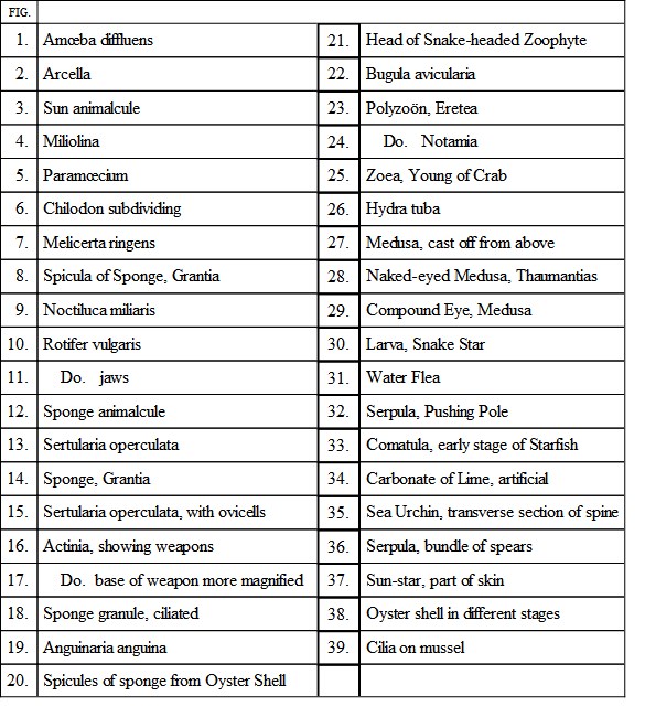
A piece of stick may be coated with a white layer, feeling rough to the touch, and full of small holes. The chances are that this will be a piece of fresh-water sponge, Spongilla fluviatílis, and by dark-field illumination particles may be seen to enter at some orifices and be ejected at others. With a very high power and a very thin section, properly prepared, these holes will be seen to be the mouths of channels which are lined by the most delicate organisms possible, each having a minute body crowned with a tiny crystal cup, in the middle of which is a long cilium, or flagellum, as it is here called (Plate XIII. Fig. 1). The currents are produced by the combined action of these flagella. In point of fact, the sponge is a colony of minute animals working harmoniously for the common good. If the specimen be found in winter the sponge will be full of tiny balls, the “gemmules” of the next season’s growth. The roughness is due to the flinty spicules, which are at once the scaffolding and the protection of the sponge, and by boiling the sponge in a mixture of nitric acid and water (half and half) these spicules will be set free, and may be washed, allowed to settle, washed again, dried, and mounted in balsam. The gemmules are coated by very beautiful spicules, consisting of two wheels connected by a rod. These may be treated in the same way. The life-history of the common sponge is as yet but imperfectly known.
Perhaps the lowest form of life is the Amœba, shown in Plate IX. Fig. 1, a mere lump of jelly, which flows along, and when it comes into contact with any likely subject for digestion flows round it, encloses it, absorbs what it can from it, and leaves it behind. A near relative of the Amœba is the Arcella (Fig. 2), which is simply an Amœba with a shell. Being unable to swim, these organisms are naturally to be most often found at the bottom of the collecting bottle, and it is always advisable to take up a portion of the débris with a dipping tube, which is then held upright on a slide with the finger upon it until the dirt settles on to the slide, when it is removed, a cover-glass put upon the dirt, and a quarter-inch power used for examination. Many forms will be discovered in this way which would otherwise escape observation.
Another curious organism, of great size (comparatively) and extreme beauty, is the sun animalcule (Actínophrys), which has a round body and long tentacles (Fig. 3), to which free-swimming organisms adhere, and by the combined action of the neighbouring ones are drawn to the body and received into it; one cannot say swallowed.
Fig. 6, Plate IX., shows the curious arithmetical process whereby the Infusoria multiply by division, a groove appearing at one point, rapidly deepening, and finally separating the animal completely into two. The species is the Chílodon, a flattened creature, ciliated all over, having a set of teeth arranged in the form of a tube, and at its fore-part a kind of membranous lip. A similar phenomenon, in an earlier stage, is shown in Fig. 26, Plate XIII., the organism in this case being Euplótes.
It has been said that sponges are colonies of extremely minute organisms, each furnished with a membranous collar or funnel, the whole looking like an exquisite wine-glass without a foot. These organisms are not always grouped in colonies, however. Many are free-growing, and may be found attached to the stems of water-plants, but they are extremely minute, and will hardly be noticed until the microscopist has acquired considerable experience, nor even then—with such an instrument as we have postulated—will he see more than a tiny pear, with a straight line, the margin of the cup, on each side of its summit. The flagellum will be quite invisible.