полная версия
Common Objects of the Microscope
We now pass from the petal of the flower to the pollen, that coloured dust, generally yellow or white, which is found upon the stamens, and which is very plentiful in many flowers, such as the lily and the hollyhock.
This substance is found only upon the stamens or anthers of full-blown flowers (the anthers being the male organs), and is intended for the purpose of enabling the female portion of the flower to produce fertile seeds. In form the pollen grains are wonderfully diverse, affording an endless variety of beautiful shapes. In some cases the exterior is smooth and marked only with minute dots, but in many instances the outer wall of the pollen grain is covered with spikes, or decorated with stripes or belts. A few examples of the commonest forms of pollen will be found on Plate III.
Fig. 17 is the pollen of the snowdrop, which, as will be seen, is covered with dots and marked with a definite slit along its length. The dots are simply tubercles in the outer coat of the grain, and are presumed to be formed for the purpose of strengthening the membrane, otherwise too delicate, upon the same principle which gives to “corrugated” iron such strength in proportion to the amount of material. Fig. 18 is the pollen of the wall-flower, shown in two views, and having many of the same characteristics as that of the snowdrop. Fig. 19 is the pollen of the willow-herb, and is here given as an illustration of the manner in which the pollen aids in the germination of plants.
In order to understand its action, we must first examine its structure.
All pollen-grains are furnished with some means by which their contents when thoroughly ripened can be expelled. In some cases this end is accomplished by sundry little holes called pores; in others, certain tiny lids are pushed up by the contained matter; and in some, as in the present instance, the walls are thinned in certain places so as to yield to the internal pressure.
When a ripe pollen-grain falls upon the stigma of a flower, it immediately begins to swell, and seems to “sprout” like a potato in a damp cellar, sending out a slender “pollen-tube” from one or other of the apertures already mentioned. In Fig. 19 a pollen-tube is seen issuing from one of the projections, and illustrates the process better than can be achieved by mere verbal description. The pollen-tubes insinuate themselves between the cells of the stigmas, and, continually elongating, worm their way down the “style” until they come in contact with the “ovules.” By very careful dissection of a fertilised stigma, the beautiful sight of the pollen-tubes winding along the tissues of the style may be observed under a high power of the microscope.
The pollen-tube is nothing more than the interior coat of the grain, very much developed, and filled with a substance technically named “fovilla,” composed of “protoplasm” (the semi-liquid substance which is found in the interior of cells), very minute starch grains, and some apparently oily globules.
In order to examine the structure of the pollen-grains properly, they should be examined under various circumstances—some dry, others placed in water to which a little sugar has been added, others in oil, and it will often be found useful to try the effect of different acids upon them.
Fig. 20 is the pollen of the common violet, and is easily recognisable by its peculiar shape and markings. Fig. 21 is the pollen of the musk-plant, and is notable for the curious mode in which its surface is belted with wide and deep bands, running spirally round the circumference. Fig. 22 exhibits the pollen of the apple, and Fig. 23 affords a very curious example of the raised markings upon the surface of the dandelion pollen. In Fig. 24 there are also some very wonderful markings, but they are disposed after a different fashion, forming a sort of network upon the surface, and leaving several large free spaces between the meshes. The pollen of the lily is shown in Fig. 25, and is a good example of a pollen-grain covered with the minute dottings which have already been described.
Figs. 26 and 27 show two varieties of compound pollen, found in two species of heath. These compound pollen-grains are not of unfrequent occurrence, and are accounted for in the following manner.
The pollen is formed in certain cavities within the anthers, by means of the continual subdivision of the “parent-cells” from which it is developed. In many cases the form of the grain is clearly owing to the direction in which these cells have divided, but there is no great certainty on this subject. It will be seen, therefore, that if the process of subdivision be suddenly arrested, the grains will be found adhering to each other in groups of greater or smaller size, according to the character of the species and the amount of subdivision that has taken place. The reader must, however, bear in mind that the whole subject is as yet rather obscure, and that further discovery may throw doubt on many theories which at present are accepted as established.
Fig. 28 shows the pollen of the furze, in which are seen the longitudinal slits and the numerous dots on the surface; and Fig. 29 is the curiously shaped pollen of the tulip. The two large yellow globular figures at each side of the Plate represent the pollen of two common flowers; Fig. 36 being that of the crocus, and Fig. 37 a pollen-grain of the hollyhock. As may be seen from the illustration, the latter is of considerable size, and is covered with very numerous projections. These serve to raise the grain from a level surface, over which it rolls with a surprising ease of motion, so much so indeed that if a little of this substance be placed on a slide and a piece of thin glass laid over it, the glass slips off as soon as it is in the least inclined, and forces the observer to fix it with paper or cement before he can place it on the inclined stage of the microscope. The little projections have a very curious effect under a high power, and require careful focusing to observe them properly; for the diameter of the grain is so large that the focus must be altered to suit each individual projection. Their office is, probably, to aid in fertilisation.
The seeds of plants are even easier of examination than the pollen, and in most cases require nothing but a pocket lens and a needle for making out their general structure. The smaller seeds, however, must be placed under the microscope, many of them exhibiting very curious forms. The external coat of seeds is often of great interest, and needs to be dissected off before it can be rightly examined. The simplest plan in such a case is to boil the seed well, press it while still warm into a plate of wax, and then dissect with a pair of needles, forceps, and scissors under water. Many seeds may also be mounted in cells as dry objects, after being thoroughly dried themselves.
A few examples of the seeds of common plants are given at the bottom of Plate III.
Fig. 38 exhibits the fruit, popularly called the seed, of the common goosegrass, or Galium, which is remarkable for the array of hooklets with which it is covered. Immediately above the figure may be seen a drawing of one of the hooks much magnified, showing its sharp curve (Fig. 39). It is worthy of remark that the hook is not a simple curved hair, but a structure composed of a number of cells terminating in a hook.
Fig. 40 shows the seed, or rather the fruit, of the common red valerian, and is introduced for the purpose of showing its plumed extremity, which acts as a parachute, and causes it to be carried about by the wind until it meets with a proper resting-place. It is also notable for the series of strong longitudinal ribs which support its external structure. On Fig. 41 is shown a portion of one of the parachute hairs much more magnified.
The seed of the common dandelion, so dear to children in their play-hours, when they amuse themselves by puffing at the white plumy globes which tip the ripe dandelion flower-stalks, is a very interesting object even to their parents, on account of its beautiful structure, and the wonderful way in which it is adapted to the place which it fills. Fig. 45 represents the seed portion of one of these objects, together with a part of the parachute stem, the remainder of that appendage being shown lying across the broken stem.
The shape of the seed is not unlike that of the valerian, but it is easily distinguished from that object by the series of sharp spikes which fringe its upper end, and which serve to anchor the seed firmly as soon as it touches the ground. From this end of the seed proceeds a long slender shaft, crowned at its summit by a radiating plume of delicate hairs, each of which is plentifully jagged on its surface, as may be seen in Fig. 46, which shows a small portion of one of these hairs greatly magnified. These jagged points are evidently intended to serve the same purpose as the spikes below, and to arrest the progress of the seed as soon as it has found a convenient spot.
Fig. 42 is the seed of the foxglove, and Fig. 43 the seed of the sunspurge, or milkwort. Fig. 47 shows the seed of the yellow snapdragon; remarkable for the membranous wing with which the seed is surrounded, and which is composed of cells with partially spiral markings. When viewed edgewise, it looks something like Saturn with his ring, or, to use a more homely but perhaps a more intelligible simile, like a marble set in the middle of a penny. Fig. 48 is a seed of mullein, covered with net-like markings on its external surface. These are probably to increase the strength of the external coat, and are generally found in the more minute seeds.
On Fig. 50 is shown a seed of the burr-reed; a structure which is remarkable for the extraordinary projection of the four outer ribs, and their powerful armature of reverted barbs. Fig. 51 shows another form of parachute seed, found in the willow-herb, where the parachute is not expanded nearly so widely as that of the valerian; neither is it set upon a long slender stem like that of the dandelion, but proceeds at once from the top of the seed, widening towards the extremity, and having a very comet-like appearance. Two more seeds only remain, Fig. 49 being the seed of Robin Hood, and the other, Fig. 52, that of the muskmallow, being given in consequence of the thick coat of hairs with which it is covered.
Many seeds can be well examined when mounted in Canada balsam.
CHAPTER VI
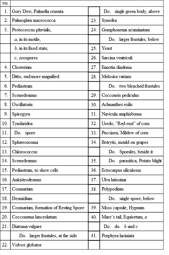
Algæ and their Growth—Desmidiaceæ, where found—Diatoms, their Flinty Deposit—Volvox—Mould, Blight, and Mildew—Mosses and Ferns—Mare’s-Tail and the Spores—Common Sea-weeds and their Growth.
On Plate IV. will be seen many examples of the curious vegetables called respectively algæ and fungi, which exhibit some of the lowest forms of vegetable life, and are remarkable for their almost universal presence in all parts of this globe, and also almost all conditions of cold, heat, or climate. Many of them are well known under the popular name of sea-weeds, others are equally familiar under the titles of “mould,” “blight,” or “mildew,” while many of the minuter kinds exhibit such capability of motion, and such apparent symptoms of volition, that they have long been described as microscopic animalcules, and thought to belong to the animal rather than to the vegetable kingdoms.
Fig. 1 represents one of the very lowest forms of vegetable life, being known to the man of science as the Palmella, and to the general public as “gory dew.” It may be seen on almost any damp wall, extending in red patches of various sizes, looking just as if some blood had been dashed on the wall, and allowed to dry there. With a tolerably powerful lens this substance can be resolved into the exceedingly minute cells depicted in the figure. Generally, these cells are single, but in many instances they are double, owing to the process of subdivision by which the plant grows, if such a term may be used.
Fig. 2 affords an example of another very low form of vegetable, the Palmoglæa, that green slimy substance which is so common on damp stones. When placed under the microscope, this plant is resolvable into a multitude of green cells, each being surrounded with a kind of gelatinous substance. The mode of growth of this plant is very simple. A line appears across one of the cells, and after a while it assumes a kind of hour-glass aspect, as if a string had been tied tightly round its middle. By degrees the cell fairly divides into two parts, and then each part becomes surrounded with its own layer of gelatine, so as to form two separate cells, placed end to end.
One of the figures, that on the right hand, represents the various processes of “conjugation,” i.e. the union and fusion together of two cells. Each cell throws out a little projection; these meet together, and then uniting, form a sort of isthmus connecting the two main bodies. This rapidly widens, until the two cells become fused into one large body. The whole subject of conjugation is very interesting, and is treated at great length in the Micrographic Dictionary of Messrs. Griffith and Henfrey, a work to which the reader is referred for further information on many of the subjects that, in this small work, can receive but a very hasty treatment.
Few persons would suppose that the slug-like object on Fig. 3, the little rounded globules with a pair of hair-like appendages, and the round disc with a dark centre, are only different forms of the same organism. Such, however, is the case, and these are three of the modifications which the Protococcus undergoes. This vegetable may be seen floating like green froth on the surface of rain-water.
On collecting some of this froth and putting it under the microscope, it is seen to consist of a vast number of little green bodies, moving briskly about in all directions, and guiding their course with such apparent exercise of volition that they might very readily be taken for animals. It may be noticed that the colour of the plant is sometimes red, and in that state it has been called the Hæmatococcus.
The “still” state of this plant is shown in the round disc. After a while the interior substance splits into two portions; these again subdivide, and the process is repeated until sixteen or thirty-two cells become developed out of the single parent-cell. These little ones then escape, and, being furnished with two long “cilia” or thread-like appendages, whirl themselves merrily through the water. When they have spent some time in this state, growing all the while, they lose their cilia, become clothed with a strong envelope, and pass into the still stage from which they had previously emerged. This curious process is repeated in endless succession, and causes a very rapid growth of the plant. The moving bodies are technically called zoospores, or living spores, and are found in many other plants besides those of the lowest order.
IV.

IV.
On Fig. 13 is delineated a very minute plant, called from its colour Chlorococcus. It may be found upon tree-trunks, walls, etc., in the form of green dust, and has recently been found to take part in forming the first stage of lichens.
A large and interesting family of the “confervoid algæ,” as these low forms of vegetable life are termed, is the Desmidiaceæ, called in more common parlance desmids. A few examples of this family are given in Plate IV.
They may be found in water, always preferring the cleanest and the brightest pools, mostly congregating in masses of green film at the bottom of the water, or investing the stems of plants. Their removal is not very easy, but is best accomplished by very carefully taking up this green slippery substance in a spoon, and straining the water away through fine muslin. They may also be separated by allowing a ring, covered with muslin, to float upon the surface of the water collected in a jar, for, being great lovers of light, they assemble where it is most abundant. An opaque jar should be used. For preservation, glycerine-gelatine seems to be the best fluid. A very full and accurate description of these plants may be found in Ralfs’ British Desmidieæ.
Fig. 4 represents one of the species of Closterium, more than twenty of which are known. These beautiful objects can be obtained from the bottom of almost every clear pool, and are of some interest on account of the circulating currents that may be seen within the living plants. A high power is required to see this phenomenon clearly. The Closteria are reproduced in various ways. Mostly they divide across the centre, being joined for a while by two half-cells. Sometimes they reproduce by means of conjugation, the process being almost entirely conducted on the convex sides. Fig. 5 represents the end of a Closterium, much magnified in order to show the actively moving bodies contained within it.
Fig. 16 is a supposed desmid, called Ankistrodesmus, and presumed to be an earlier stage of Closterium.
Fig. 6 is a very pretty desmid called the Pediastrum, and valuable to the microscopist as exhibiting a curious mode of reproduction. The figure shows a perfect plant composed of a number of cells arranged systematically in a star-like shape; Fig. 15 is the same species without the colouring matter, in order to show the shape of the cells. The Pediastrum reproduces by continual subdivision of the contents of each cell into a number of smaller cells, termed “gonidia” on account of their function on the perpetuation of the species. When a sufficient number has been formed, they burst through the envelope of the original cell, taking with them a portion of its internal layer, so as to form a vesicle, in which they move actively. In a few minutes they arrange themselves in a circle, and after a while they gradually assume the perfect form, the whole process occupying about two days. Fig. 18 exhibits an example of the genus Desmidium. In this genus the cells are either square or triangular in their form, having two teeth at their angles, and twisted regularly throughout their length, causing the wavy or oblique lines which distinguish them. The plants of this genus are common, and may be found almost in any water. I may as well mention that I have obtained nearly all the preceding species, together with many others, from a little pond on Blackheath.
Fig. 7 is another desmid called Scenedesmus, in which the cells are arranged in rows of from two to ten in number, the cell at each extremity being often furnished with a pair of bristle-like appendages. Fig. 14 is another species of the same plant, and both may be found in the water supplied for drinking in London, as well as in any pond.
A common species of desmid is seen at Fig. 12, called Sphærozosma, looking much like a row of stomata set chainwise together. It multiplies by self-division.
Fig. 17 is a specimen of desmid named Cosmarium, plentifully found in ponds on heaths and commons, and having a very pretty appearance in the microscope, with its glittering green centre and beautifully transparent envelope. The manner in which the Cosmarium conjugates is very remarkable, and is shown at Fig. 19.
The two conjugating cells become very deeply cleft, and by degrees separate, suffering the contents to pour out freely, and, as at present appears, without any envelope to protect them. The mass, however, soon acquires an envelope of its own, and by degrees assumes a dark reddish-brown tint. It is now termed a sporangium, and is covered with a vast number of projections, which in this genus are forked at their tip, but in others, which also form sporangia, are simply pointed. The Closteria conjugate after a somewhat similar manner, and it is not unfrequent to find a pair in this condition, but in their case the sporangium is quite smooth on its surface.
Another very remarkable family of confervoid algæ is that which is known under the name of Oscillatoriæ, from the oscillating movement of the plant. They are always long and filamentous in character, and may be seen moving up and down with a curious irregularity of motion. Their growth is extremely rapid, and may be watched under a tolerably powerful lens, thus giving many valuable hints as to the mode by which these plants are reproduced. One of the commonest species is represented at Fig. 8.
Figs. 9, 10, and 11 are examples of another family, called technically the Zygnemaceæ, because they are so constantly yoked together by conjugation. They all consist of a series of cylindrical cells, set end to end, and having their green contents arranged in similar patterns. Two of the most common and typical species are here given.
Fig. 9 is the Spirogyra, so called from the spiral arrangement of the chlorophyll; and Fig. 10 is the Tyndaridea, or Zygnema, as it is called by some writers. A casual inspection will show how easy it is to distinguish the one from the other. Fig. 11 represents a portion of the Tyndaridea during the process of conjugation, showing the tube of connection between the cells and one of the spores.
We now arrive at the diatoms, so called because of their method of reproduction, in which it appears as if a cut were made right along the original cell. The commonest of these plants is the Diatóma vulgáre, seen in Fig. 21 as it appears while growing. The reproduction of this plant is effected by splitting down the centre, each half increasing to the full size of the original cell; and in almost every specimen of water taken from a pond, examples of this diatom undergoing the process of division will be distinguished. It also grows by conjugation. The diatoms are remarkable for the delicate shell or flinty matter which forms the cell skeleton, and which will retain its shape even after intense heat and the action of nitric acid. While the diatoms are alive, swimming through the water, their beautiful markings are clearly distinct, glittering as if the form were spun from crystalline glass. Just above the figure, and to the right hand, are two outlines of single cells of this diatom, the one showing the front view and the other the profile.
Fig. 20 is an example of a diatom—Cocconéma lanceolátum—furnished with a stalk. The left-hand branch sustains a “frustule” exhibiting the front view, while the other is seen sideways.
Another common diatom is shown in Fig. 23, and is known by the name of Synedra. This constitutes a very large genus, containing about seventy known species. In this genus the frustules are at first arranged upon a sort of cushion, but in course of time they mostly break away from their attachment. In some species they radiate in every direction from the cushion, like the spikes of the ancient cavalier’s mace.
Fig. 24 is another stalked diatom called Gomphonéma acuminátum, found commonly in ponds and ditches. There are nearly forty species belonging to this genus. A pair of frustules are also shown which exhibit the beautiful flinty outline without the coloured contents (technically called endochrome).
Fig. 27 is a side view of a beautiful diatom, called Eunótia diadéma from its diadem-like form. There are many species of this genus. When seen upon the upper surface, it looks at first sight like a mere row of cells with a band running along them; but by careful arrangement of the light its true form may easily be made out.
Fig. 28 represents a very common fresh-water diatom, named Melosíra várians. The plants of this genus look like a cylindrical rod composed of a variable number of segments, mostly cylindrical, but sometimes disc-shaped or rounded. An end view of one of the frustules is seen at the left hand, still coloured with its dots of “endochrome,” and showing the cylindrical shape. Immediately above is a figure of another frustule seen under both aspects with the endochrome removed.
A rather curious species of diatom, called Cocconeïs pedículus, is seen at Fig. 29 as it appears on the surface of common water-cress. Sometimes the frustules, which in all cases are single, are crowded very closely upon each other and almost wholly hide the substance on which they repose. Fig. 30 is another diatom of a flag-like shape, named Achnanthes, having a long slender filament attached to one end of the lower frustule, representing the flag-staff. There are many wonderful species of such diatoms, some running almost end to end like a bundle of sticks, and therefore called Bacillária; others spreading out like a number of fans, such as the genus Licmophora; while some assume a beautiful wheel-like aspect, of which the genus Meridion affords an excellent example.